
|
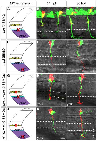
Fig. 9 Netrins can prevent extension of a second CaP axon into ventral muscle.
Projected confocal stacks of living dye-labeled motoneurons in embryos injected with various combinations of MOs. A, D, G, J. Cartoons of a trunk spinal hemisegment detailing ntn1a, ntn1b, and ntn2 mRNA expression. The red X indicates which netrin transcript was knocked down in adjacent panels. B–C. ntn1b SBMO-injected Tg(mnx1:GFP) embryo showing motoneurons (green) and rhodamine dextran-labeled VaPs (yellow). At 24 (B) and 36 hpf (C) VaP axons are stopped at muscle pioneers (upper dashed line). E, F, H, I, K, L. Embryos with individually-labeled CaP and VaP pairs. Dashed ellipses outline motoneuron cell bodies. In some cases interneurons (*) were also injected with dye during the labeling procedure. E–F. In ntn2 SBMO-injected embryos the VaP axon (green) stalled at the muscle pioneers while the CaP axon (red) extended into ventral muscle at 24 hpf (E) and at 36 hpf (F). H–I. In ntn1a plus ntn1b SBMO-injected embryos two axons extended into ventral muscle at 24 hpf (H), but by 36 hpf (I) one axon retracted to the level of the second intermediate target (lower dashed line), becoming CaP-like (green). K–L. In the absence of ntn1a and ntn2 only one axon (green) initially extended to the third intermediate target (K). However, by 36 hpf a second axon (red) extended to the third intermediate target, thus both cells become CaPs (L). Dashed ellipses in L outline the motoneuron cell bodies. Scale bar = 20 μm.