
|
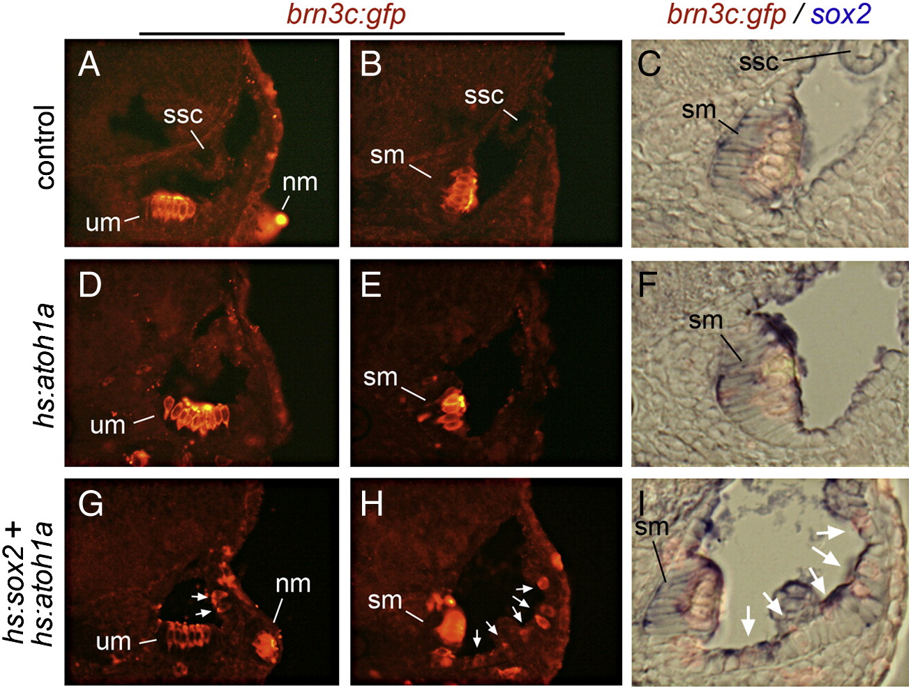
Fig. 7 Sox2 expands sensory competence at later stages. Transverse sections showing otic expression of brn3c:gfp (red) and sox2 (blue) in a control embryo (A–C), a hs:atoh1a transgenic embryo (D–F) and a hs:atoh1a;hs:sox2 double transgenic embryo (G–I). Embryos were serially heat shocked at 45 and 48 hpf and fixed at 72 hpf for staining and sectioning. Shown are sections passing through the anterior end (A, D, G) or the posterior end (B, C, E, F, H, I) of the otic vesicle. Positions of the utricular macula (um), saccular macula (sm), semicircular canals (ssc) and lateral line neuromasts (nm) are indicated. White arrows (G–I) mark ectopic hair cells. Specimens in C, F, and I are enlargements of images B, E, and H, respectively and are shown in brightfield with fluorescence to clarify the spatial relationship between hair cells and sox2 expression.
Reprinted from Developmental Biology, 358(1), Sweet, E.M., Vemaraju, S., and Riley, B.B., Sox2 and Fgf interact with Atoh1 to promote sensory competence throughout the zebrafish inner ear, 113-21, Copyright (2011) with permission from Elsevier. Full text @ Dev. Biol.