
|
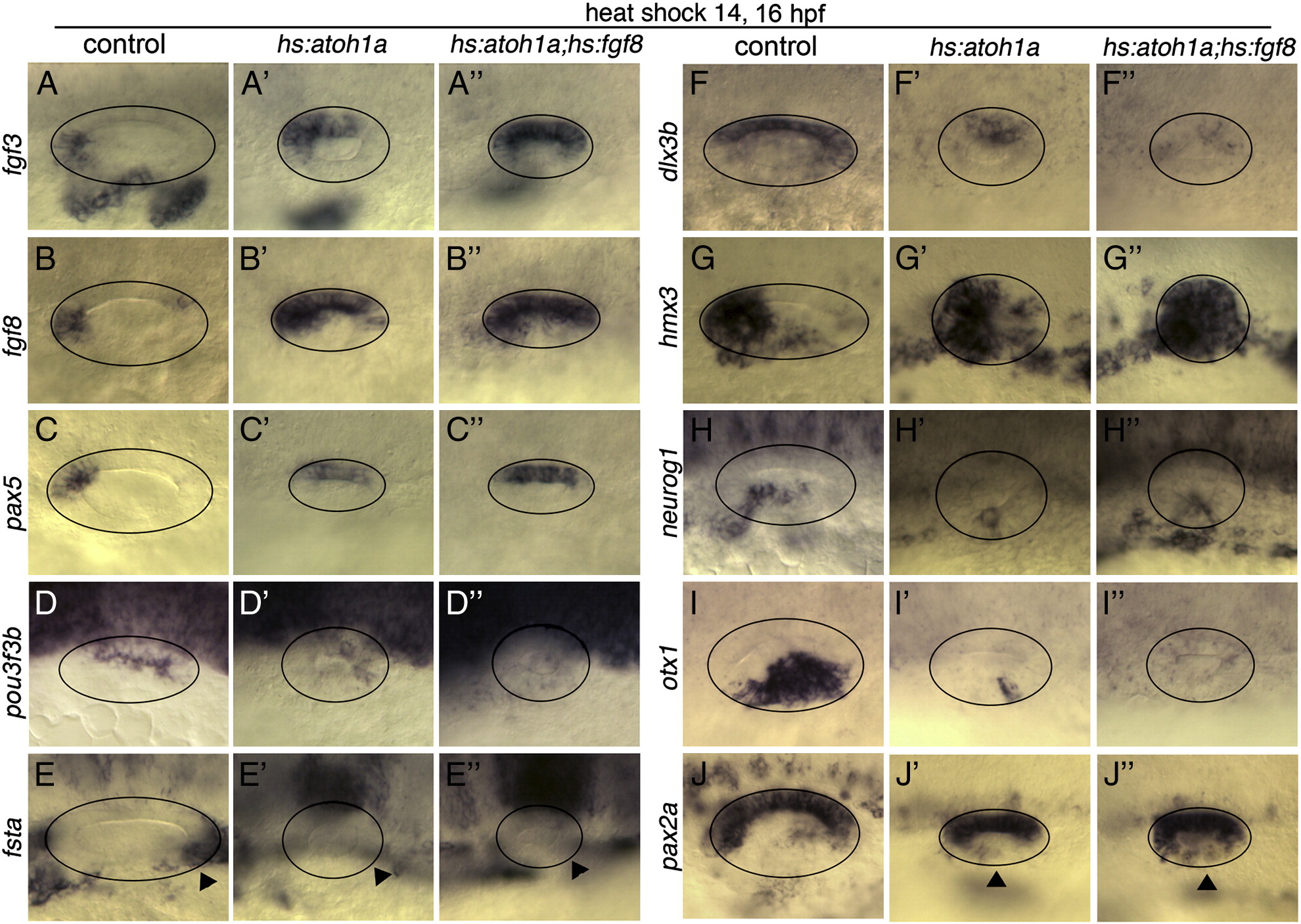
Fig. 6 Axial patterning following co-activation of hs:atoh1a and hs:fgf8. Expression of various otic markers in control embryos (A–J), hs:atoh1a transgenic embryos (A′–J′) and hs:atoh1a;hs:fgf8 double transgenic embryos (A′′–J′′). Embryos were serially heat shocked at 14 and 16 hpf and fixed for processing at 26 hpf. Images show dorsal views (A–C′′) or dorsolateral views (D–J′′), with anterior to the left. Circles outline the otic vesicle. Arrowheads in E–E′′ mark expected location of fsta in the posterior otic vesicle. Arrowheads in J–J′′ indicate expanded domains of pax2a in the lateral wall of the otic vesicle.
Reprinted from Developmental Biology, 358(1), Sweet, E.M., Vemaraju, S., and Riley, B.B., Sox2 and Fgf interact with Atoh1 to promote sensory competence throughout the zebrafish inner ear, 113-21, Copyright (2011) with permission from Elsevier. Full text @ Dev. Biol.