
Image
|
Figure Caption
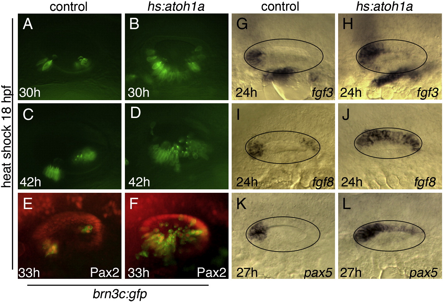
Fig. 2 Otic vesicle patterning following hs:atoh1a activation at 18 hpf. (A–F) Expression of brn3c:gfp (green) in the utricle and saccule of control embryos (A, C, E) and in hs:atoh1a transgenic embryos (B, D, F) at the indicated times. (E, F) Co-staining with anti-Pax2 in red. (G–L) Otic expression of fgf3, fgf8, and pax5 in control embryos (G, I, K) and expanded expression in hs:atoh1a transgenic embryos (H, J, L). All images show dorsolateral views with anterior to the left and dorsal up (A–H) or dorsal views with anterior to the left and medial up (I–L).
Acknowledgments
This image is the copyrighted work of the attributed author or publisher, and
ZFIN has permission only to display this image to its users.
Additional permissions should be obtained from the applicable author or publisher of the image.
Reprinted from Developmental Biology, 358(1), Sweet, E.M., Vemaraju, S., and Riley, B.B., Sox2 and Fgf interact with Atoh1 to promote sensory competence throughout the zebrafish inner ear, 113-21, Copyright (2011) with permission from Elsevier. Full text @ Dev. Biol.