
|
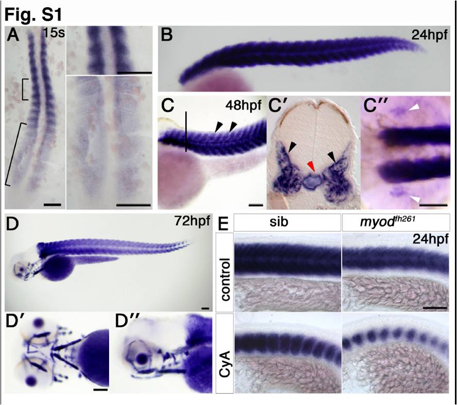
Fig. S1 Expression of miR-206 in zebrafish development. In situ RNA hybridization for miR-206. Dorsal (A) or lateral (E) flatmounts, lateral (B, C, D, D′′), dorsal (C′′) or ventral (D′) wholemounts, anterior to top (A) or left (B–E). A. Expression is detected late in adaxial slow fibre differentiation and early in fast myogenesis. B. At 24 hpf, expression is restricted to the differentiated somitic muscle. C. By 48 hpf, expression is abundant in somitic fast muscle (arrowheads). A transverse section at the level indicated by the line in C, shows signal in somitic fast muscle (C′, black arrowheads). Notochord signal (red arrow) is detected in some embryos. Pectoral fin muscle expression is also detected (C′′, white arrowheads). D. At 72 hpf, the forming cranial muscles also contain miR-206. E. miR-206 expression is reduced in somitic muscle of myodfh261 mutant and further reduced after cyclopamine treatment, indicating that miR-206 is expressed in slow muscle fibres. Bars = 100 μm.
Reprinted from Developmental Biology, 358(1), Hinits, Y., Williams, V.C., Sweetman, D., Donn, T.M., Ma, T.P., Moens, C.B., and Hughes, S.M., Defective cranial skeletal development, larval lethality and haploinsufficiency in Myod mutant zebrafish, 102-12, Copyright (2011) with permission from Elsevier. Full text @ Dev. Biol.