
|
Fig. 3
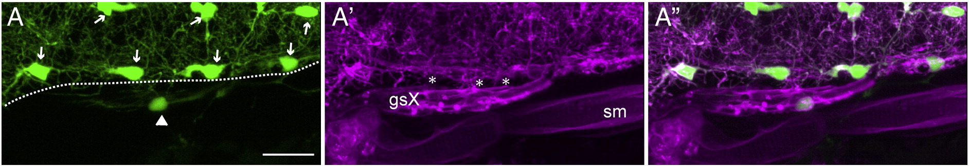
Sox:rfp+ peripheral glia contribute to the hindbrain transition zone of the Xth nerve. Images shown are from a 4 dpf olig2:gfp;sox:rfp larva (n = 3). Panel A is a dorsal view (anterior at left) of one side of the hindbrain, caudal to the otocyst, showing GFP+ oligodendrocytes and their processes. Arrows point to oligodendrocyte cell bodies and an arrowhead indicates an olig2-expressing cell present outside of the hindbrain. The dashed line marks the approximate position of the hindbrain boundary. A′ shows membrane-targeted RFP in the peripheral glia composing the vagal sheath, as well as in some CNS oligodendrocytes and their processes. Note the RFP+ processes (marked by asterisks) that appear to link the glial sheath to the hindbrain at several points. A" is a merge of A and A′. Notice that the GFP+ cell seen in panel A is present in the glial sheath. sm, skeletal muscle; gsX, glial sheath of the Xth nerve. Scale bars are 20 μm.
Reprinted from Developmental Biology, 357(2), Cox, J.A., Lamora, A., Johnson, S.L., and Voigt, M.M., Diverse mechanisms for assembly of branchiomeric nerves, 305-17, Copyright (2011) with permission from Elsevier. Full text @ Dev. Biol.