
|
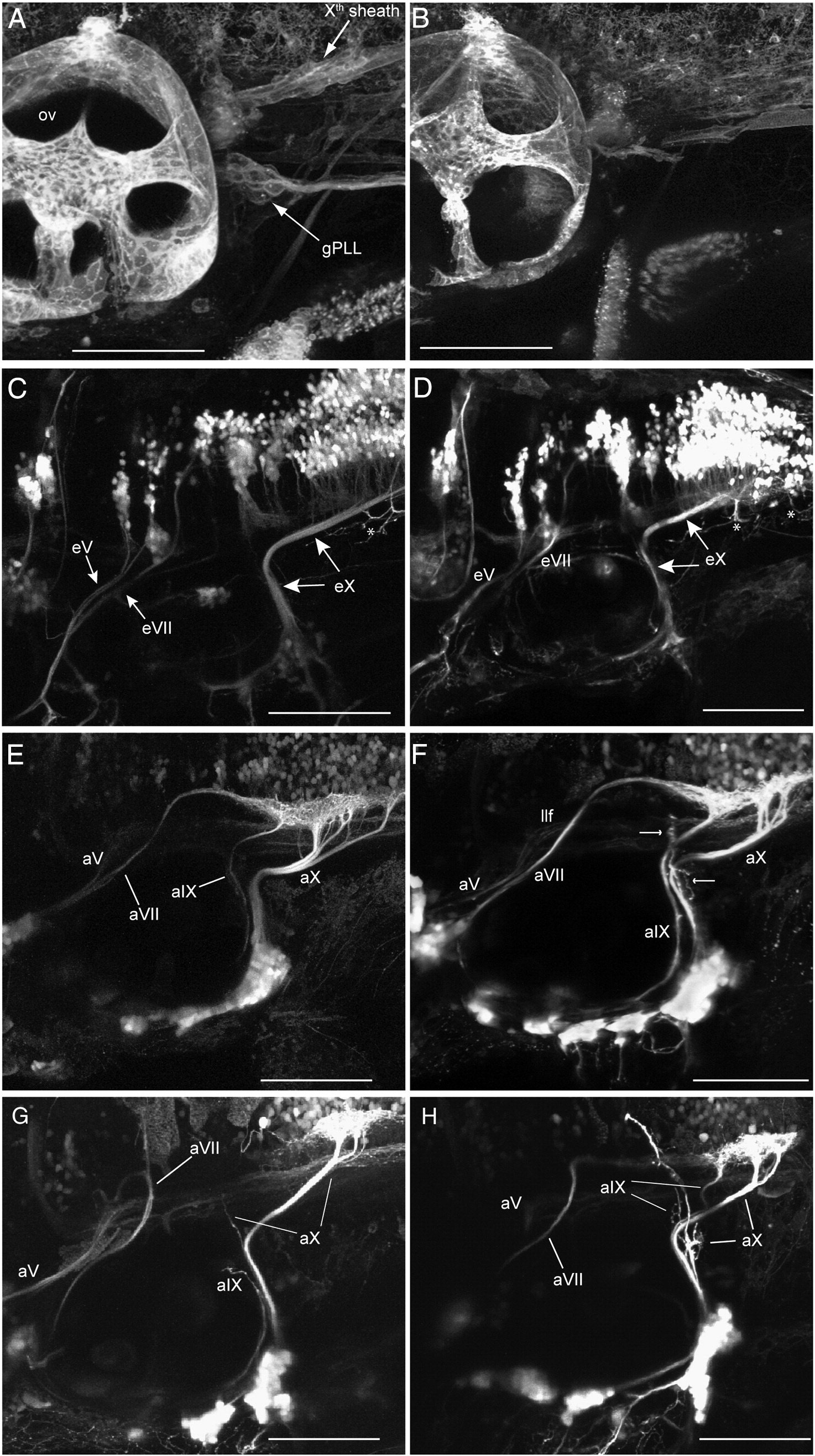
Fig. 11 foxd3 nulls have lost peripheral glia, resulting in some cranial afferents becoming defasciculated and misrouted. A: 4 dpf control foxd3+/ -sox:rfp larva, B: 4 dpf mutant foxd3 - / -;sox:rfp larva showing loss of the Xth sheath and gPLL satellite glia. C: 4 dpf control foxd3+/ -;isl:gfp larva, D: 4 dpf mutant foxd3 - / -;isl:gfp larva showing correct proximal eV, eVII and eX projections. E: 4 dpf control foxd3+/ -3.2:gfp larva, F–H: 4 dpf mutant foxd3 - / -3.2:gfp larvae show a spectrum of axon misrouting; subsets of aVII (G), aIX (G,H) and aX (F–H) afferent axons are defasciculated and misrouted (e.g., arrows in F), whereas the remaining axons project correctly. gPLL, posterior lateral line ganglion satellite cells; ov, otic vesicle; eV, efferent nerve of bmnV; eVII, efferent nerve of bmnVII; eX, efferent nerve of bmnX; aV, afferent nerve of gV; aVII, afferent nerve of gVII; aIX, afferent nerve of gIX; aX, afferent nerve of gX; llf, lateral longitudinal fascicle. Anterior to left, dorsal at top. All scale bars are 100 μm.
Reprinted from Developmental Biology, 357(2), Cox, J.A., Lamora, A., Johnson, S.L., and Voigt, M.M., Diverse mechanisms for assembly of branchiomeric nerves, 305-17, Copyright (2011) with permission from Elsevier. Full text @ Dev. Biol.