
|
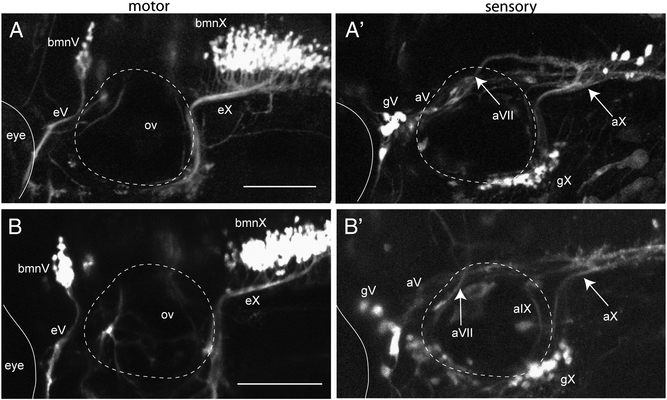
Fig. 10
Hh signaling does not have a direct role on central sensory axon pathfinding. Images are from 4 dpf larvae co-expressing isl:gfp (left panels) and 3.2.cherry (right panels). The images shown are representative of 4 control and 7 treated embryos imaged from 2 separate experiments. Panels A, A′ is a control larva (0.5% ethanol); B,B′ cycA treatment (50 μM cycA/0.5% ethanol) beginning at 24 hpf shows no effect on either sets of axons. Dotted circle is otic vesicle (ov), eyes are demarcated by thin solid line. Arrows in A′ and B′ point to vagal afferents entering hindbrain. bmnV, motor nucleus of V; bmnX, vagal motor nucleus; eV, efferent nerve of bmnV; eX, efferent nerve of bmnX; aV, afferent nerve of gV; gV, trigeminal sensory ganglion; gX, vagal sensory ganglion; aVII, afferent nerve of gVII; aIX, afferent nerve of gIX; aX, afferent nerve of gX. Anterior to left, dorsal at top. Scale bars are 100 μm.
Reprinted from Developmental Biology, 357(2), Cox, J.A., Lamora, A., Johnson, S.L., and Voigt, M.M., Diverse mechanisms for assembly of branchiomeric nerves, 305-17, Copyright (2011) with permission from Elsevier. Full text @ Dev. Biol.