
|
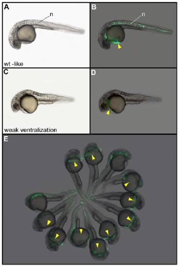
Fig. S5 Phenotype and localization of the injected clone of cells in nocodazole-treated embryos injected with Wnt8a and GFP mRNAs. After treatment with nocodazole and injection of 25 pg of Wnt8a and 100 pg of GFP mRNAs into one blastomere at the 64-cell stage, embryos displaying a wild-type–like phenotype (A and B) always express the GFP in axial structures including the hatching gland (yellow arrowhead). Embryos displaying a weak ventralization phenotype (C and D) lack the notochord but always develop a hatching gland expressing GFP. (E) Field of embryos showing that the hatching gland is always derived from the injected blastomere. The two embryos lacking the hatching gland correspond to the class of strong ventralization phenotypes for which this tissue is not formed. (A, C, and E) Bright field. (B, D, and F) Localization of GFP fluorescent cells. n, notochord. Embryos are 30 h old and in lateral view.