
|
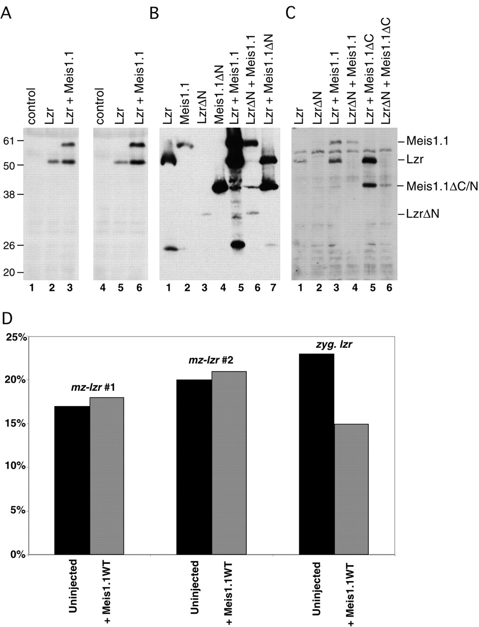
Fig. 7 Bidirectional stabilization of Meis1.1 and Lzr. (A-C) Embryos were injected with mRNAs and lysed to isolate proteins as shown. All proteins were expressed as N-terminal fusions with the Myc epitope to ensure equal rates of translation initiation and to detect proteins by immunoblot. (A) By comparing Lzr protein levels in lane 2 and 5 (Lzr alone) with levels in lane 3 and 6 (Lzr + Meis1.1WT), co-expression of Meis1.1WT increases detectable Lzr by 3.4-fold. (B) Meis1.1 levels are increased eightfold by co-expressing Lzr, in comparison to the non-binding Meis1.1ΔN (compare lanes 1, 5, and 7). Meis levels are increased similarly by co-expressed Lzr protein, but not significantly by LzrΔN (compare lanes 2, 5, and 6). (C) Meis1.1ΔC is stabilized by Lzr and can stabilize Lzr protein (compare lanes 1, 5, and 6). Apparent molecular weights in kDa are labeled and the anticipated position of each protein is indicated. (D) Expression of Meis1.1WT does not decrease the percentage of embryos from a maternal-zygotic lzr (mz-lzr) mutant clutch with abnormal krox20. However, the same Meis1.1WT mRNA injected into zygotic lzr does reduce the percentage of abnormal krox20, indicating the ability to rescue the phenotype associated with zygotic loss-of-function.