
|
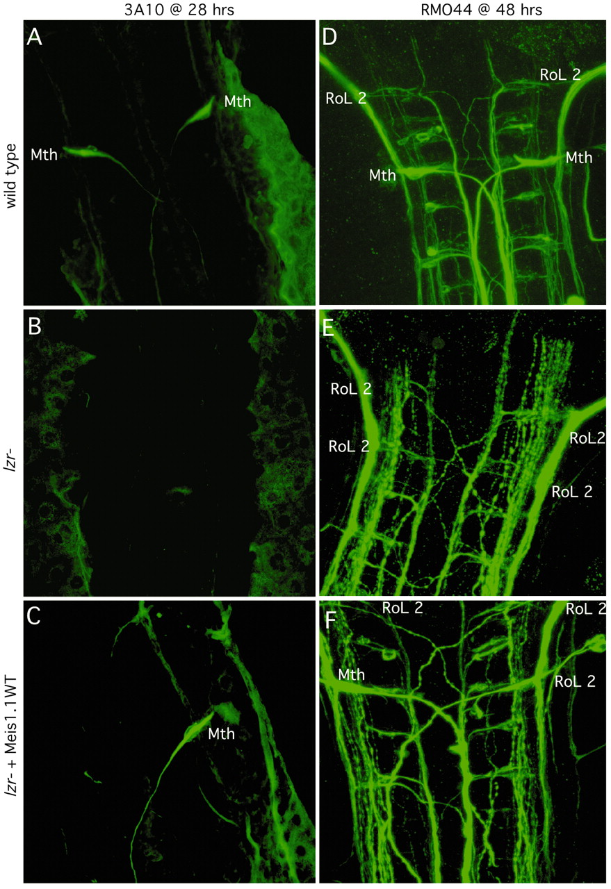
Fig. 5 Expression of meis1.1WT in lzr mutants increases the percentage of embryos with correctly specified Mauthner (labeled Mth) neurons. Each panel is oriented with anterior towards the top. (A-C) Either wild-type or lzr- embryos were injected with Meis1.1WT mRNA as shown on left. 28 hour embryos were stained with 3A10, an antibody which recognizes the Mth neuron and its axon. (D-F) 48 hour embryos, as described on the left were stained with RMO44, an antibody that recognizes the identifiable primary reticulospinal neurons of zebrafish. Neuronal cell bodies are identified and labeled as shown (RoL 2, which is normally found in r2 of a wild-type embryo; Mth, which is in r4).