
Fig. S2
- ID
- ZDB-IMAGE-111011-29
- Publication
- Fujimoto et al., 2011 - Gal80 intersectional regulation of cell-type specific expression in vertebrates
- All Figures
- Figures for Fujimoto et al., 2011
|
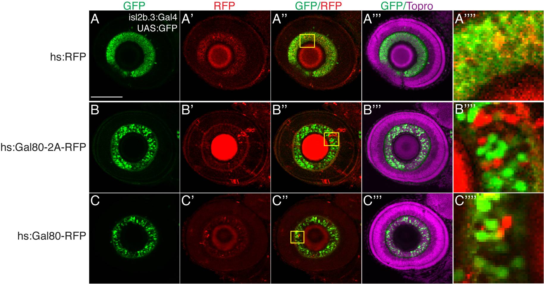
Fig. S2 Subgroups of neurons can be distinguished by inhibiting Gal4-driven expression with a fluorescently tagged Gal80. Single slice confocal images, lateral views, anterior to left, dorsal up, of 72 hours postfertilization (hpf) eyes in Tg(isl2b.3:Gal4)zc65; Tg(UAS:GFP) embryos. Immunostaining for green fluorescent protein (GFP), green; TagRFP, red; Topro3 nuclear stain, magenta. A–A′′ ′: Embryos injected with hsp70l:TagRFP (no Gal80 expression). B–B′′ ′: Embryos injected with hsp70l:Gal80-2A-TagRFP and heat-shocked at 48 hpf show expression of TagRFP (RFP) and inhibition of Gal4-dependent expression. C–C′′ ′: Embryos injected with hsp70l:Gal80-TagRFP and heat-shocked at 48 hpf show RFP expression and concomitant inhibition of Gal4-dependent expression. (A′′ ′′–C′′ ′′) shows magnified view of the insets in (A′′–C′′), demonstrating disjoint GFP and RFP expression. Scale bar = 50 μm.