
Fig. 6
- ID
- ZDB-IMAGE-111011-28
- Publication
- Fujimoto et al., 2011 - Gal80 intersectional regulation of cell-type specific expression in vertebrates
- All Figures
- Figures for Fujimoto et al., 2011
|
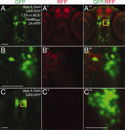
Fig. 6 Subgroups of neurons can be genetically defined by expressing Gal4 and Gal80 with partially overlapping enhancers. Confocal ventral views, anterior to top, of brain in 72 hours postfertilization (hpf) embryos. Immunostaining for green fluorescent protein (GFP), green; TagRFP, red. A–A′′: Maximum intensity projections of Tg(otpb.A:Gal4)zc67; Tg(UAS:GFP); Tg(f.TH.m:NLS-Gal80opt-2A-TRFP)zc78 embryos shows nonoverlap of Gal80 and GFP expression. B–B′′: Boxed inset area from A′′ shows single confocal slice of individual cells expressing either GFP or red fluorescent protein (RFP) but not both. C: Single confocal slice of Tg(otpb.A:Gal4)zc67; Tg(UAS:GFP) embryo and inset (C2–C3) shows full complement of cells express GFP in absence of Gal80. Scale bars = 50 μm; 25 μm in B,C.