
Fig. 5
- ID
- ZDB-IMAGE-111011-27
- Publication
- Fujimoto et al., 2011 - Gal80 intersectional regulation of cell-type specific expression in vertebrates
- All Figures
- Figures for Fujimoto et al., 2011
|
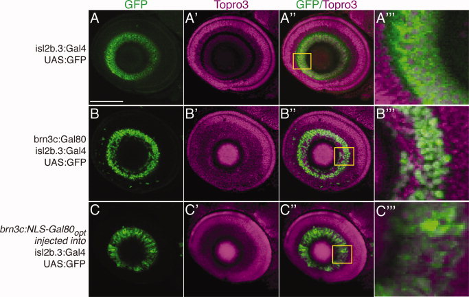
Fig. 5 Nuclear localization and codon optimization improves function of Gal80. Confocal maximum projections, lateral views, anterior to left, dorsal up, of eyes in Tg(isl2b.3:Gal4)zc65; Tg(UAS:GFP) 72 hours postfertilization (hpf) embryos. Immunostaining for green fluorescent protein (GFP), green; Topro3 nuclear stain, magenta. A–A′′ ′: Tg(isl2b.3:Gal4)zc65; Tg(UAS:GFP) transgenic embryos with no Gal80 show GFP expression in all retinal ganglion cells (RGCs). Inset and A′′ ′ shows high power magnification of single confocal slice. B–B′′ ′: Triple transgenic Tg(isl2b.3:Gal4)zc65; Tg(UAS:GFP); Tg(brn3c:Gal80) shows inhibition of Gal4-dependent GFP expression in approximately 30% of RGCs. Inset and B′′ ′ shows high power magnification of single confocal slice. C–C′′ ′: Transient injection with construct carrying “improved” Gal80 into Tg(isl2b.3:Gal4)zc65; Tg(UAS:GFP) embryos demonstrates inhibition similar to that of stable lines carrying native Gal80. Improved Gal80 has nuclear localization signal (NLS) and is codon optimized (“opt”). Scale bar = 50 μm.