
|
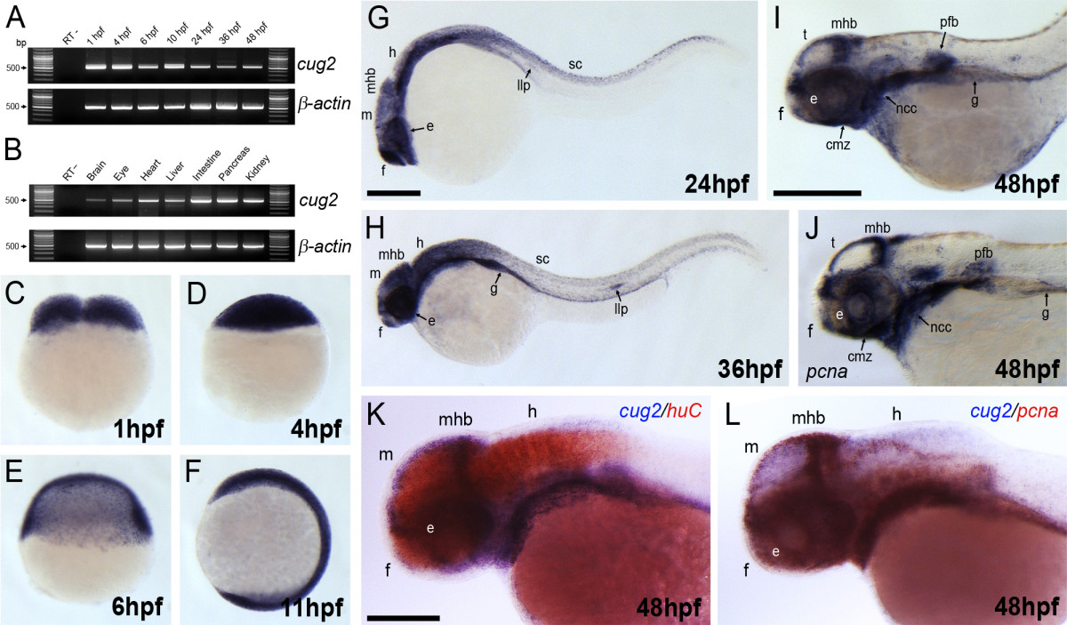
Fig. 2 Expression pattern of cug2 in developing zebrafish embryos. A. Temporal expression profile of zebrafish cug2 by RT-PCR. Zebrafish cug2 transcripts have maternal and zygotic expression. β-actin is the loading control. B. The expression of cug2 is detected in the brain, eye, heart, liver, intestine, pancreas, and kidney in adult zebrafish. C-F. In cleavage (C), blastula (D), gastrula (E), and segmentation stages (F), cug2 transcripts are ubiquitously expressed throughout the embryonic body. G. At 24 hpf, cug2 transcripts are detected in the eye (e), forebrain (f), midbrain (m), midbrain-hindbrain boundary (mhb), hindbrain (h), spinal cord (sc), and lateral line primordium (llp). H. Expression of cug2 detected in the lateral line primordium, gut (g) and CNS at 36 hpf. I. Expression of cug2 in the ciliary marginal zone (cmz) in the eyes, tectum (t), midbrain-hindbrain boundary, neural crest cells (ncc), pectoral fin buds (pfb) and gut at 48 hpf. J. Expression pattern of pcna at 48 hpf. Scale bar = 200 μm. K. At 48 hpf, expression domain of cug2 (blue) is not overlapped with that of huC (red), a differentiating neuronal marker. Scale bar = 100 μm. L. cug2-expressing region is almost overlapped with pcna-expressing proliferating zones at 48 hpf.