
Image
|
Figure Caption
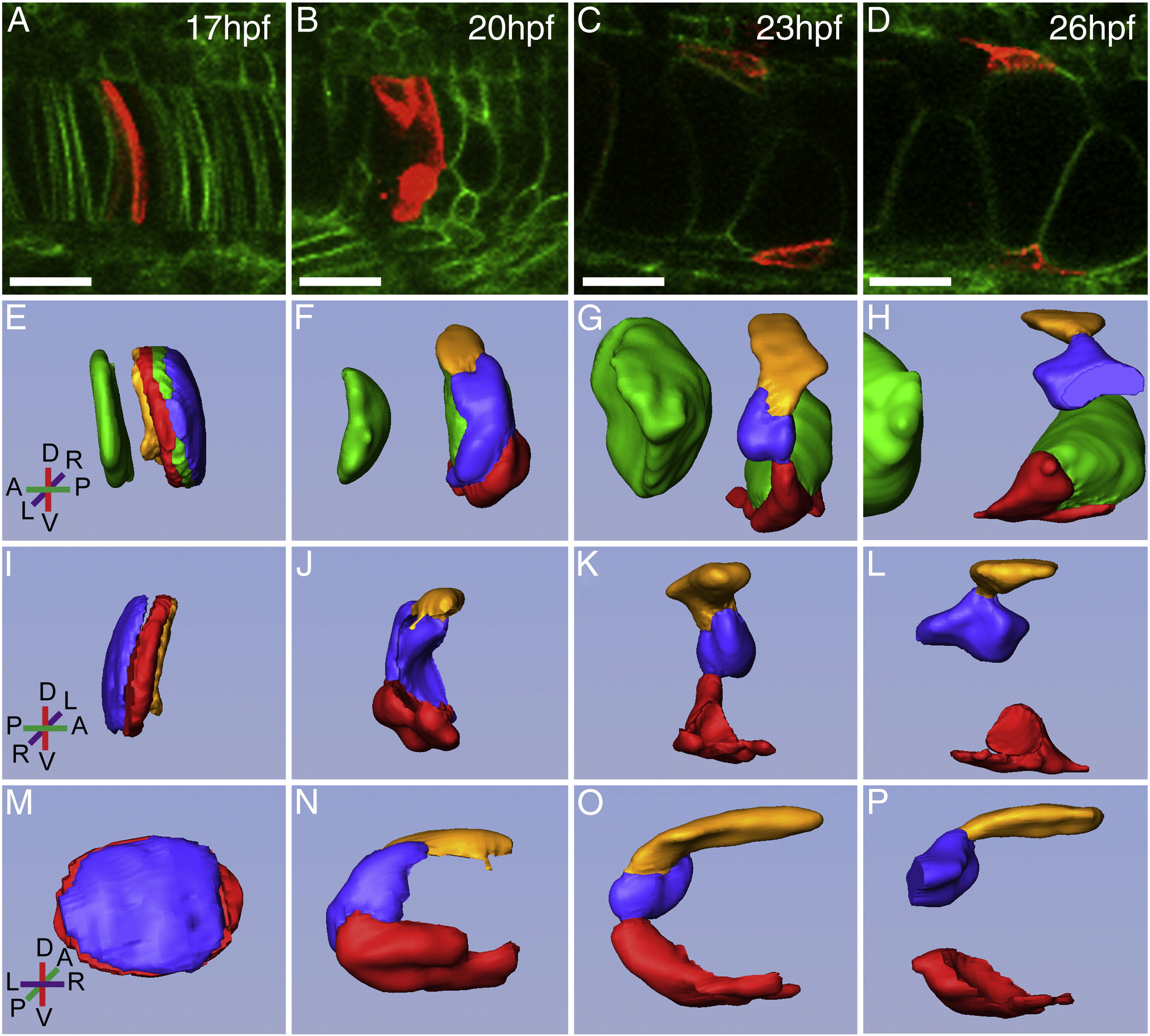
Fig. 5 Formation of the epithelial notochord sheath cells. (A–D) Confocal images of Tg(β-actin:HRAS-EGFP)vu119 fish injected with - 1.7kbcol2a1a:mCherry-CAAX plasmid to track the snapping, retraction, and spreading process of NSCs over the course of 9 h. (E–P) 3D rendering of labeled cells from confocal images. (E–H) Lateral right and I–L. left lateral view with green vacuolated cells removed. (M–P) posterior view along notochord axis. Coordinates indicate anterior/posterior (A/P), dorsal/ventral (D/V), and left/right (L/R) axes. Scale bar = 20 μm.
Acknowledgments
This image is the copyrighted work of the attributed author or publisher, and
ZFIN has permission only to display this image to its users.
Additional permissions should be obtained from the applicable author or publisher of the image.
Reprinted from Developmental Biology, 357(2), Dale, R.M., and Topczewski, J., Identification of an evolutionarily conserved regulatory element of the zebrafish col2a1a gene, 518-31, Copyright (2011) with permission from Elsevier. Full text @ Dev. Biol.