
Fig. 5
- ID
- ZDB-IMAGE-110929-31
- Genes
- Antibodies
- Publication
- Wythe et al., 2011 - Hadp1, a newly identified pleckstrin homology domain protein, is required for cardiac contractility in zebrafish
- All Figures
- Figures for Wythe et al., 2011
|
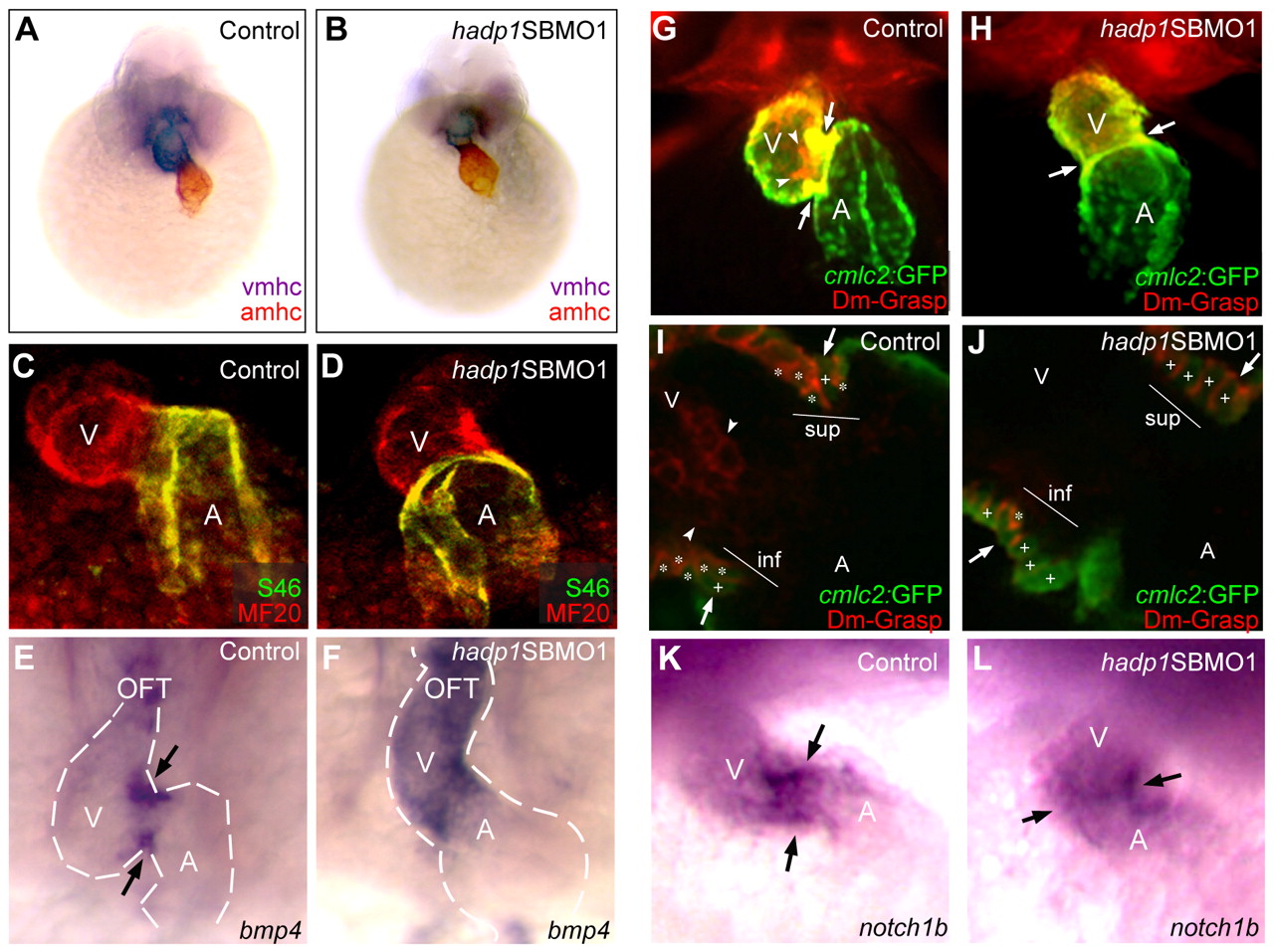
Fig. 5
Non-chamber myocardium fails to differentiate properly in hadp1 morphants. (A–J) Frontal views at 48 hpf (head to the top) illustrate expression of chamber-specific genes in control and hadp1 morphant embryos. (A,B) atrial myosin heavy chain (amhc; red; atrial marker) and ventricular myosin heavy chain (vmhc; purple; ventricular marker) were visualized by double in situ hybridization. (C,D) MF20 (red) and S46 (green) were visualized by whole mount immunofluorescence. (E,F) bmp4 expression was examined by in situ hybridization. Ventricle (V), atrium (A) and outflow tract (OFT) are indicated. Arrows indicate the AV boundary, and the dotted line demarcates the cardiac silhouette. (G,H) Immunofluorescence was used to study cmlc2:GFP (green) and DM-GRASP (red) expression. Arrows and arrowheads indicate myocardium and endocardium, respectively. (I,J) Confocal analysis of the AV canal of control (I) and hadp1 morphant (J) hearts shown in G,H. The superior (sup) and inferior (inf) aspects of the canal are shown, and remodeled trapezoidal cells are indicated with asterisks, whereas non-remodeled columnar cardiomyocytes are indicated with plus signs. Arrows and arrowheads indicate myocardium and endocardium, respectively. (K,L) notch1b expression was examined by in situ hybridization. Ventricle (V) and atrium (A) are indicated. Arrows indicate the AV boundary.