
|
Fig. 6
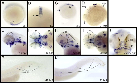
Expression patterns of lgr6 during zebrafish development. (A–K) lgr6 expression was examined by whole-mount in situ hybridization. Lateral views, anterior to the left (A, C, D, E, G, H, and K). A vegetal pole view of the tail-bud region (B). Dorsal views, anterior to the left (F and I). A ventral view of (H and I) (J). an, anterior neuromasts; ALLP, anterior lateral line primordium; ch, ceratohyals; di, diencephalon; kV, Kupffer’s vesicle; mc, Meckel’s cartilages; nc, notochord; ov, otic vesicles; pa, pharyngeal arches; pf, pectoral fin buds; pn, posterior neuromasts; PLLP, posterior lateral line primordium; tr, trabeculae.
Reprinted from Gene expression patterns : GEP, 11(7), Hirose, K., Shimoda, N., and Kikuchi, Y., Expression patterns of lgr4 and lgr6 during zebrafish development, 378-83, Copyright (2011) with permission from Elsevier. Full text @ Gene Expr. Patterns