
|
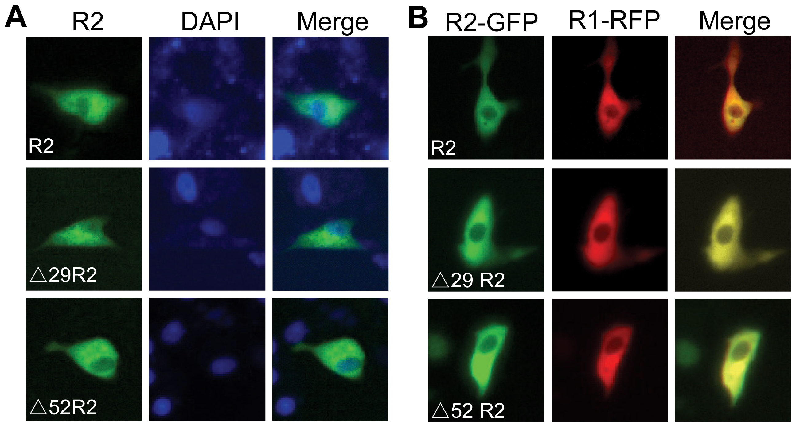
Fig. 8
Subcellular localization of zebrafish R2 isoforms.
(A) Immunofluorescence staining was performed to detect the subcellular distribution of zebrafish R2 isoforms in transfected Hela cells. Three R2 isoforms were tagged with a Flag at N-terminus. At 36 h after transfection, R2 isoforms were detected with primary anti-Flag antibody and FITC-conjugated secondary antibody. Nuclei were stained with DAPI. (B) Co-localization of fluorescent protein-tagged zebrafish R1 and R2 isoforms in the cytosol of Hela cells. R2 isoforms or R1 of zebrafish were fused with GFP or RFP to their C-termini. At 36 h after transfection, images were directly acquired under fluorescence microscopy.