
|
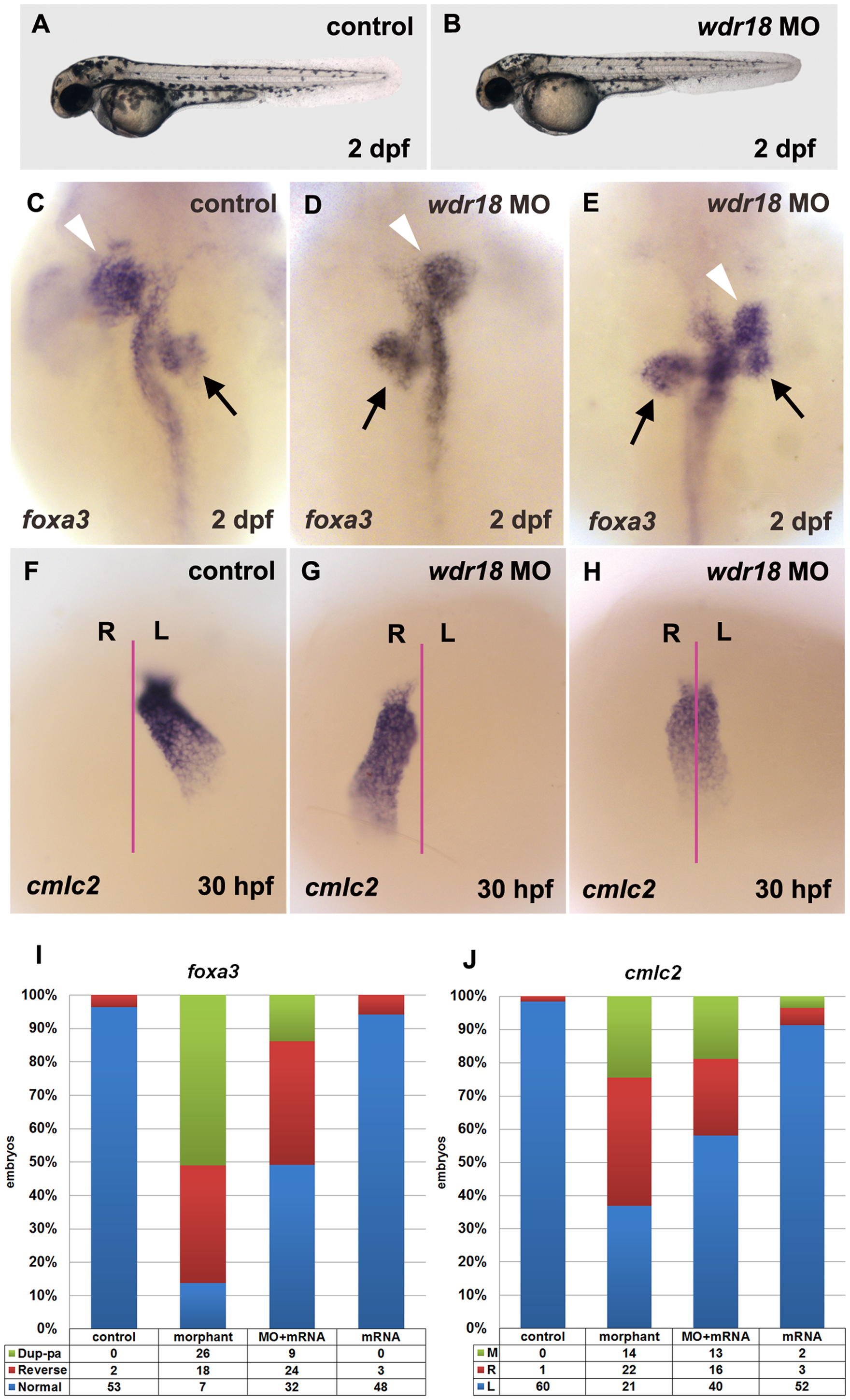
Fig. 2
Knockdown of wdr18 disturbed L-R asymmetry development of internal organs.
(A, B) The overall appearance of a wdr18 morphant embryo (B) is comparable to a wildtype control embryo (A). (C) Expression of foxa3, a marker for the endoderm, showed that the liver (white arrowheads) and pancreas (black arrows) were positioned to the left and the right of the midline in a control embryo at 2 dpf, respectively. In morphant embryos, the positions of the liver and pancreas were reversed (D), or the pancreas appeared duplicated (two black arrows) when the liver was randomized (E). cmlc2, a marker for the heart, was examined in embryos at 30 hpf. After the heart tube formation, leftward bending of the heart was observed in control embryos (F). By contrast, wdr18 morphant embryos exhibited randomized movements: leftward, rightward (G), or no movement (H). The proportion of each phenotype is summarized in (I) and (J). Dup-pa in (I) indicates embryos with duplicated pancreas. M, R, and L in (J) stand for middle, right, and left, respectively. Injection of 100 pg wdr18 mRNA partially rescued the endodermal organ phenotype. The statistical results are summarized in (I) and (J). (C–E) Dorsal views, anterior to the top. (F–H) Ventral views, anterior to the top.