
|
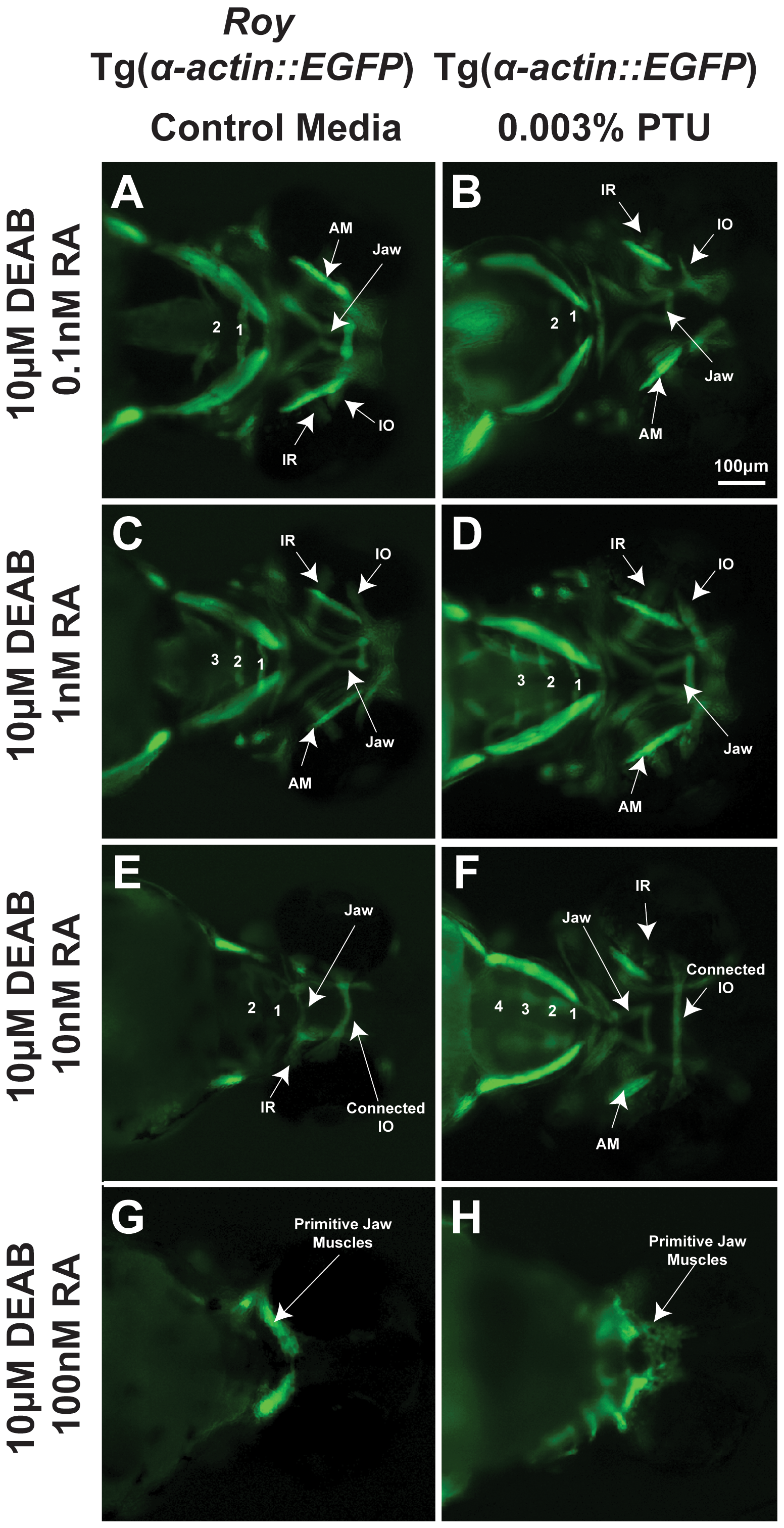
Fig. 2
PTU alters sensitivity of craniofacial tissues to retinoic acid.
72 hpf Tg(α-actin::EGFP) embryos (ventral view) were treated with 10 μM DEAB at 24 hpf and increasing concentrations of retinoic acid (RA; 0.1 nM, 1 nM, 10 nM, 100 nM) at 28 hpf in the roy background (in the absence of PTU; A, C, E, G) or presence of 0.003% PTU added at 12 hpf (B, D, F, H). In the roy background and in the absence of PTU, 1 nM retinoic acid improved the DEAB-induced effects on pharyngeal arch formation (C), but at higher concentrations (10 nM (E) and 100 nM (G)), retinoic acid suppressed pharyngeal arch development. DEAB-induced extraocular muscle disorganization in the presence of PTU (Figure 1A, B) was rescued by 0.1 nM retinoic acid (B). The DEAB effects on pharyngeal arch development in the presence of PTU was improved by 1 nM (D) and rescued by 10 nM retinoic acid (F). Teratogenic effects of retinoic acid on jaw musculature and pharyngeal arches were lessened in the presence of PTU (F, H compared to E, G). IR, inferior rectus; IO, inferior oblique; AM, anterior mandibulae.