
Image
|
Figure Caption
Fig. S2
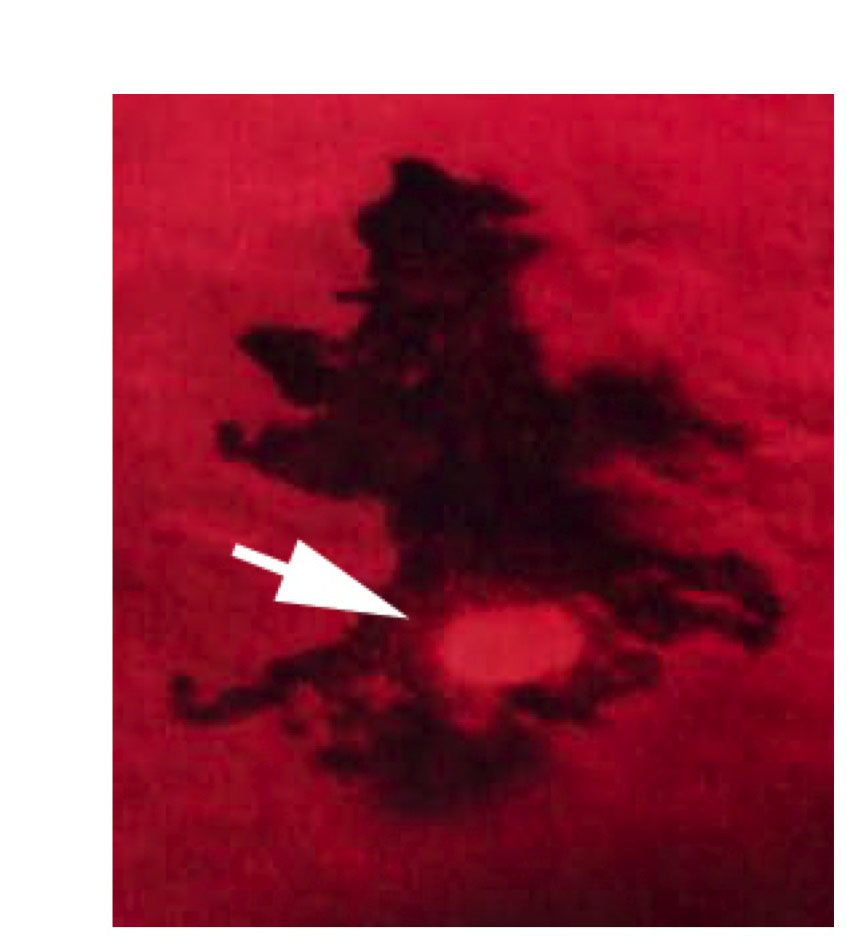
Pigmented embryonic melanocytes of trunk and tail are positive for the proliferative antigen phospho-Histone H3 (p-H3). p-H3-positive nucleus on dorsal yolk of 36 hpf embryo. p-H3-positive melanocytes were found at low frequencies in 28-36 hpf embryos sampled (2.0±2.7 cells per embryo, n=30). p-H3-expressing cells were found widely distributed throughout the embryo, and are likely to contribute to all of the embryonic melanocyte stripes.
Acknowledgments
This image is the copyrighted work of the attributed author or publisher, and
ZFIN has permission only to display this image to its users.
Additional permissions should be obtained from the applicable author or publisher of the image.
Full text @ Development