
|
Fig. 10
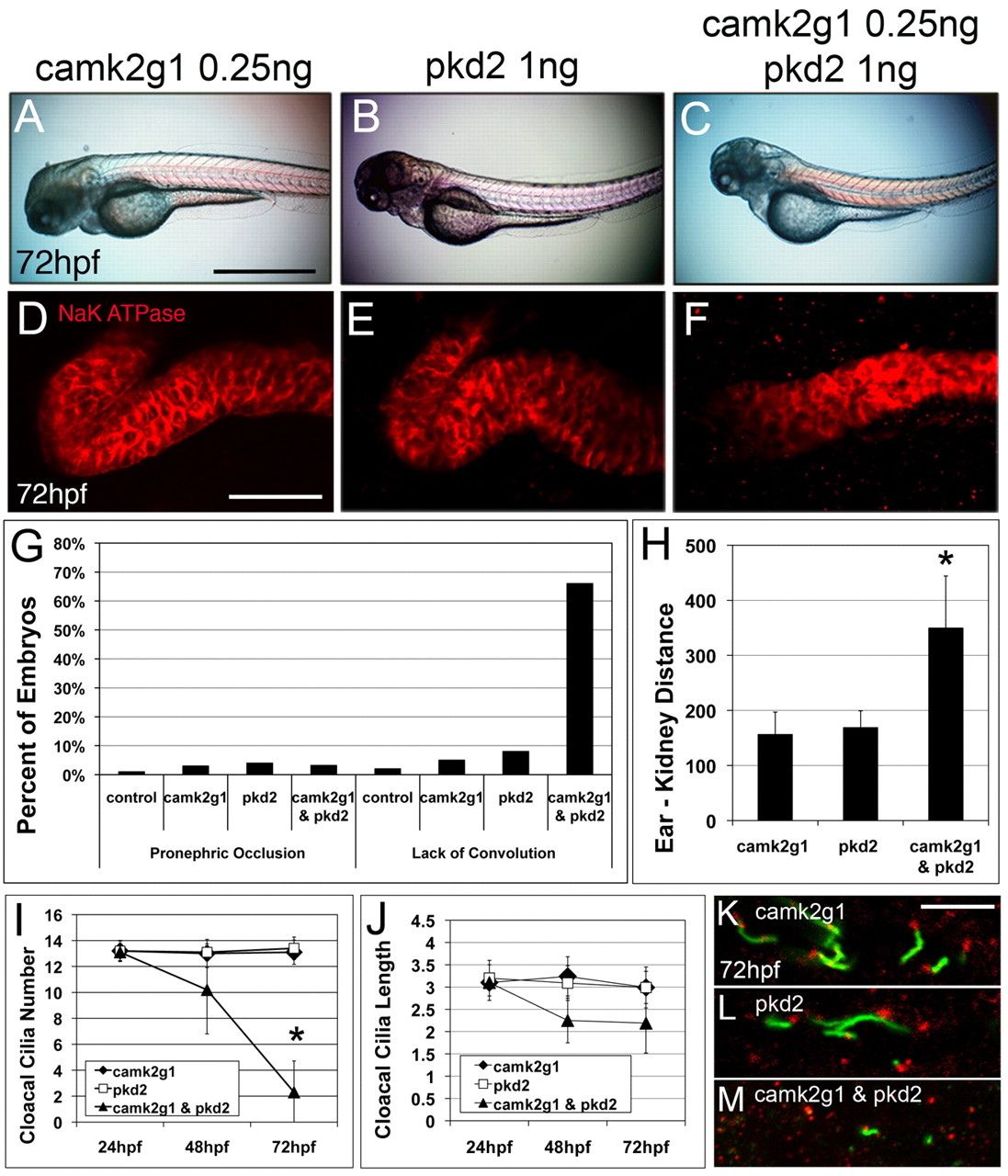
CaMK-II and PKD2 act in the same molecular pathway. (A-F) Embryos injected with camk2g1 MO (0.25 ng), pkd2 MO (1 ng) or camk2g1 and pkd2 MOs (0.25 ng and 1 ng, respectively) were imaged at 72 hpf from lateral perspectives using DIC optics (A-C) or dorsal perspectives using immunofluorescence microscopy to identify α1 Na+/K+-ATPase (D-F). (G,H) Pronephric occlusions and convolution at 72 hpf (G; n=80-125) and pronephric migration as assessed by posterior ear-anterior kidney distance in µm at 72 hpf (H; n=60-82). (I,J) Cloacal cilia number (I) and cilia length (J) were determined at 24, 48 and 72 hpf. *P<0.005. (K-M) 72 hpf morphants immunolabeled with acetylated tubulin (green) and γ–tubulin (red) show a loss of cloacal cilia but retention of the basal body. Scale bars: 500 μm in A; 20 μm in D; 5 μm in K.