
|
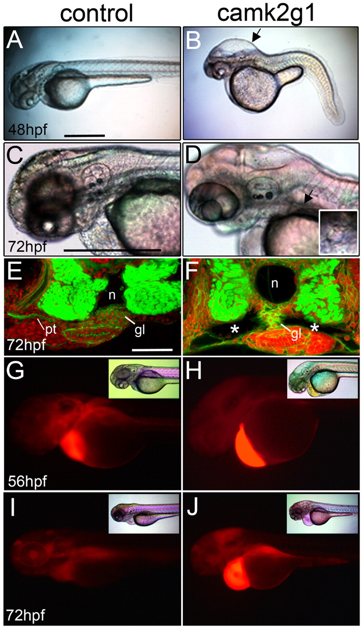
Fig. 4
Suppression of γ1 CaMK-II (camk2g1) induces cyst formation. (A,B) Lateral images of 48 hpf morphants injected with the camk2g1 MO (1 ng) but not the control MO (1 ng) show hydrocephaly (arrow) and axis compression. (C,D) Hydrocephaly and cysts are visible at 72 hpf in camk2g1 morphants (inset: dorsal view of anterior cyst (indicated by the arrow). (E,F) Cysts (*) in camk2g1 morphants are revealed in vibratome sections stained with Alexa-Fluor-488-phalloidin and propidium iodide; pt, pronephric tubules; n, notochord; gl, glomerulus. (G-J) Renal filtration was observed in control embryos but not morphant embryos. Rhodamine-dextran was injected into the pericardial region at 56 hpf and then embryos were imaged with DIC optics (inset) or for fluorescence within 20 minutes (56 hpf) and then again at 72 hpf. Scale bars: 500 μm in A,C; 50 μm in E.