
|
Fig. 6
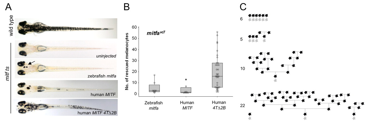
Human MITF 4TΔ2B promotes differentiated cell division. (A) Images of zebrafish embryos (day 5 postfertilization). Single-cell mitfavc7 embryos were injected with zebrafish mitfa, human MITF or human MITF4TΔ2B expressed from the mitfa promoter and grown at 30°C for 5 days. Uninjected mitfavc7 embryos were used as a control to monitor endogenous melanocyte development; no melanocytes developed in mitfavc embryos at 30°C (n=50). (B) Box plot of a representative experiment showing the range of melanocytes on individual zebrafish expressing zebrafish mitfa (n=8), human MITF (n=8) or human MITF4TΔ2B (n=35) from the mitfa promoter in mitfavc7 mutants grown at 30°C. Each data set has been repeated at least three times. Significance is observed within the dataset (P=0.001), analysis of variance (ANOVA). Post-hoc analysis shows significance between zebrafish mitfa and human MITF4TΔ2B; 95% confidence interval; 14.42 (2.03, 26.81), and between human MITF and MITF4TΔ2B; 95% CI; 16.04 (3.65, 28.43). (C) Schematic representation of melanocyte cell lineage analysis (as described in Fig. 5) for four embryos expressing MITF4TΔ2B. Total final number of melanocytes in imaged region is indicated.