
|
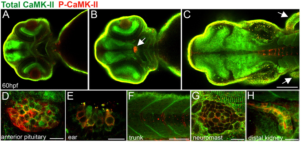
Fig. 2
Localization of activated and total CaMK-II in 60 hpf zebrafish embryos. (A-D) Dorsal views of zebrafish brain optical sections (from ventral to dorsal) showing CaMK-II in the forebrain (A), optic nerve and retina and the anterior pituitary (B, arrow, D) and, as expected, in the midbrain, hindbrain (C) and fins (C, arrows). As expected, total CaMK-II is found in the midbrain, hindbrain and fins. (E-F) Lateral views reveal P-CaMK-II in the hair cells of the ear (E), sarcomeres and somite boundaries (F,G), neuromasts (G) and kidney (H). Scale bars: in C, 10 μm for A,B; 10 μm in D,E,G,H; 100 μm in C; 50 μm in F.