
|
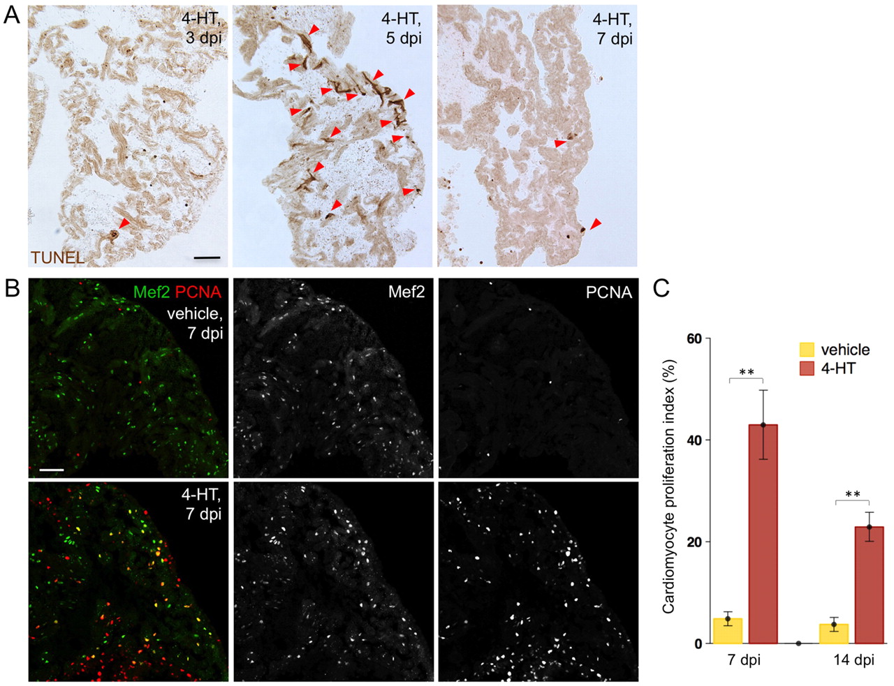
Fig. 5
Atrial cardiomyocyte ablation and regeneration. (A) TUNEL staining of atrial sections from Z-CAT fish injected with 4-HT. Arrowheads indicate TUNEL-positive muscle. (B) Cardiomyocyte proliferation at 7 days post-injection (dpi) in atrial sections from Z-CAT animals injected with vehicle or 4-HT, assessed by Mef2 and PCNA staining. 4-HT-injected animals display widespread atrial PCNA+ cardiomyocytes. Scale bars: 50 μm. (C) Quantification of atrial cardiomyocyte proliferation in Z-CAT fish injected with vehicle or 4-HT, at 7 and 14 dpi. For each group, six to eight animals were assessed. Mean±s.e.m. **P<0.005, Student′s t-test.