
|
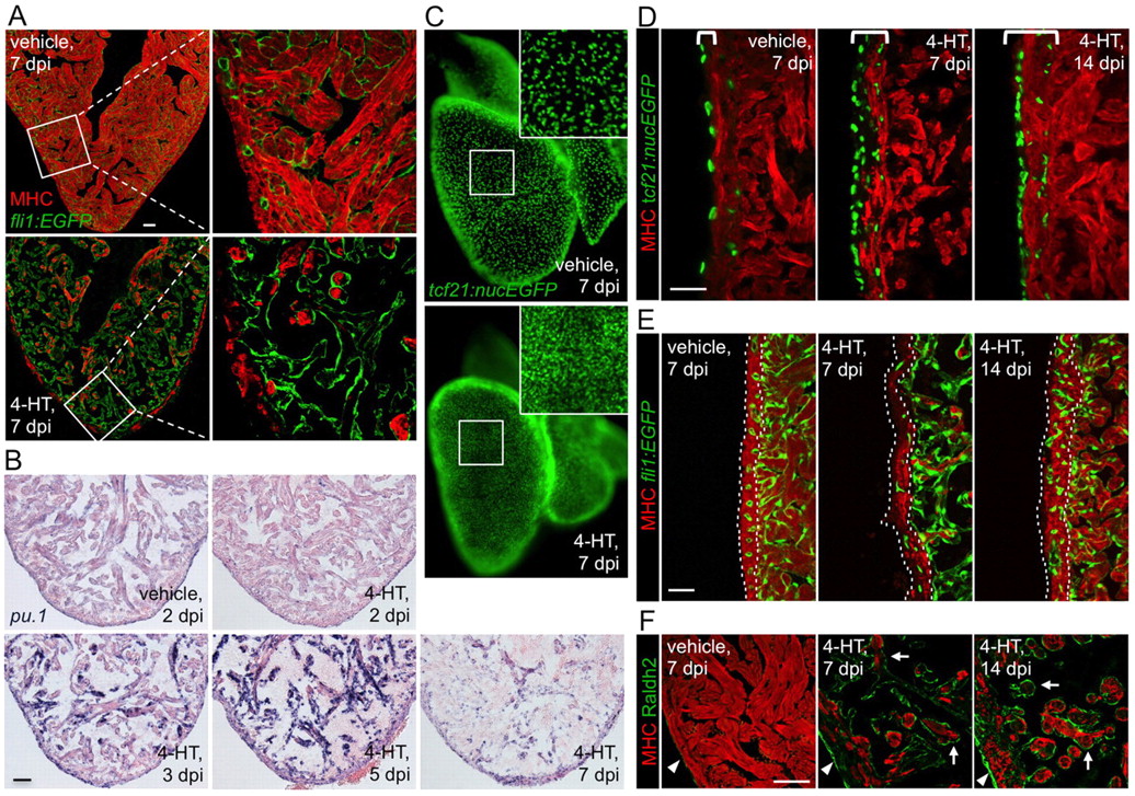
Fig. 4
Effects of cardiomyocyte ablation on non-muscle cells. (A) Visualization of endocardial cells (green) and muscle (red) in sections from vehicle-or 4-HT-injected Z-CAT fish. The endocardial layer appears intact one week after massive cardiomyocyte loss. Single confocal slices are shown. (B) In situ hybridization for pu.1-expressing cells, which are prominent by 3-5 dpi. (C) Whole-mount visualization of epicardial nuclei in vehicle- or 4-HT-injected Z-CAT fish at 7 dpi, indicating higher epicardial cell density after myocardial injury. Inset shows enlarged view of boxed area. (D) Histological visualization of epicardial cells after myocardial ablation. Epicardial cells proliferate by 7 dpi and are incorporated into the regenerating myocardial wall by 14 dpi. Brackets indicate area containing tcf21-expressing epicardial cells. (E) Visualization of coronary vascular endothelial cells (green) in sections of the ventricular wall (outlined by dotted lines). Coronary vasculature is reduced in 4-HT-injected Z-CAT animals by 7 dpi, but restored by 14 dpi. (F) Sections from Z-CAT ventricles stained for the retinoic acid (RA)-synthesizing enzyme Raldh2. RA synthesis appears low in vehicle-injected tissue, but is strongly induced in epicardial (arrowheads) and endocardial (arrows) structures after cardiomyocyte ablation. Single confocal slices are shown. Scale bars: 50 μm.