
|
Fig. 3
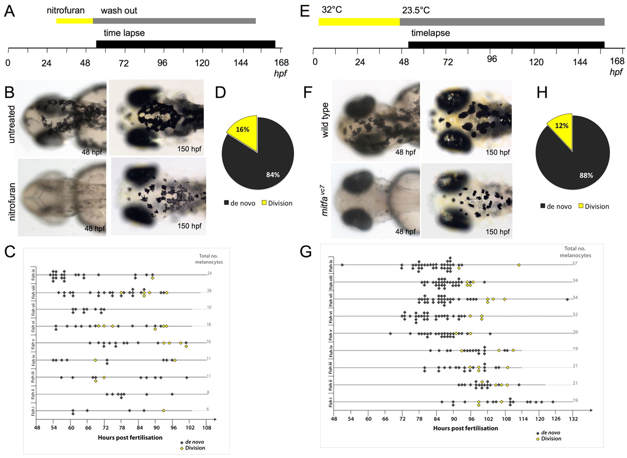
Melanocyte division events are enhanced during melanocyte regeneration. (A) Workflow of nitrofuran treatment from <30 hpf to 50 hpf to promote loss of differentiated melanocytes. After washout (grey bar), the embryos were immobilized in agarose and imaged by time-lapse microscopy (black bar). (B) Images of control and nitrofuran-treated embryos at the time of embedding in agarose and at the end of the time-lapse imaging. (C,D) Quantitative analysis of melanocytes derived de novo from unpigmented cells (grey diamonds; n=111) and from division of pigmented cells (yellow diamonds; n=21) in nine different zebrafish (i-ix). (E) Workflow of mitfavc7 temperature-sensitive experimental conditions. Zebrafish embryos with the mitfavc7 mutation grown at 32°C from 4 hpf until <48 hpf (yellow bar) do not develop ontogenic melanocytes. Upon shifting to the permissive conditions (grey bar), the embryos are immobilized in agarose and imaged by time-lapse microscopy (black bar). (F) Images of wild type and mitfavc7 embryos at the time of embedding in agarose and at the end of the time-lapse imaging. No neural crest-derived melanocytes are visible at 48 hpf (although non-neural crest pigmented cells in the eye remain pigmented) and melanocytes have repopulated the animal by 150 hpf. (G,H) Quantitative analysis of melanocytes derived de novo from unpigmented cells (grey diamonds; n=214) and from division of pigmented cells (yellow diamonds; n=29) in nine individual zebrafish (i-ix).