
|
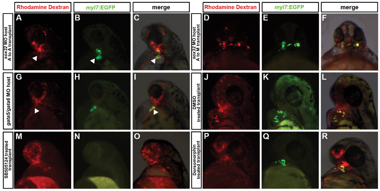
Fig. 8
Effects of host tissues and signaling pathways on the ability of cBAF-expressing cells to form myocardial cells. (A-R) Transgenic myl7:EGFP donor embryos were injected with tetramethylrhodamine dextran and gata5/smarcd3b RNA, and transplanted at 4 hpf into the animal pole of host embryos injected with morpholinos targeting sox32 (A-F), and gata5 and gata6 (G-I). (J-R) Donor cells as per above were transplanted into the animal pole of wild-type host embryos, with transplants treated from sphere stage (4 hpf) onwards with DMSO [as drug treatment control (J-L)], 40 μM SB-505124 (M-O) or 10 μM dorsomorphin (P-R). In A-F, cells were transplanted from the animal pole (A) to the host margin (M) or animal pole (A) as indicated. Embryos are shown at 48 hpf. (A-C,G-R) Lateral views with anterior towards the top of images; (D-F) ventral views with anterior towards the top.