
Fig. 4
- ID
- ZDB-IMAGE-110907-24
- Publication
- Lou et al., 2011 - Smarcd3b and Gata5 promote a cardiac progenitor fate in the zebrafish embryo
- All Figures
- Figures for Lou et al., 2011
|
Fig. 4
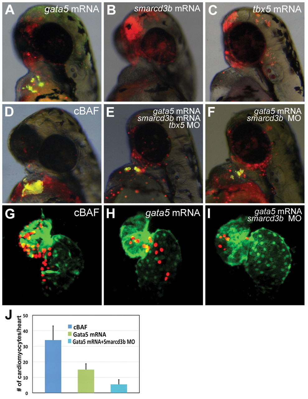
Role of single components of cBAF in promotion of cardiomyocyte differentiation. (A-F) Transgenic myl7:EGFP donor embryos were injected with tetramethylrhodamine dextran (red) and gata5 mRNA (A), smarcd3b mRNA (B), tbx5 mRNA (C), gata5/smarcd3b mRNA (D), gata5/smarcd3b mRNA and tbx5 morpholino (E), or gata5 mRNA and smarcd3b morpholino (F). At 4 hpf, donor cells were transplanted to the animal pole of wild-type hosts. Donor cell cardiomyocyte differentiation was assessed at 48 hpf by EGFP fluorescence. All images are overlays of red channel, green channel and bright field, such that donor cell-derived cardiomyocytes appear yellow. (G-J) Transgenic myl7:dsRedExp-nuc donor embryos were injected with gata5/smarcd3b RNA, gata5 RNA alone or gata5 RNA in conjunction with smarcd3b morpholino. Transplantation was carried out as above. At 48 hpf, donor-derived cardiomyocytes were quantified via nuclear-localized dsRed signal. (J) Results shown as number of donor cardiomyocytes per positive transplant embryo (mean±s.e.m., P<0.05).