
|
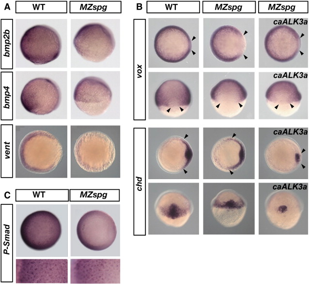
Fig. 4
BMP signaling and expression of zygotic BMP pathway genes in MZspg. (A) Expression of BMP pathway genes is reduced in MZspg embryos. From top to bottom: Whole-mount in-situ hybridization of wild-type (WT) embryos and MZspg mutants showing expression of bmp2b, bmp4 (lateral view, dorsal to the right, 75% epiboly) and vent (animal view, dorsal to the right, shield stage). (B) Constitutively active BMP receptor 1a (caALK3a) can rescue dorsalization of MZspg embryos at midgastrulation. Whole-mount in situ hybridization of wild-type (WT) embryos, MZspg mutants, and MZspg mutants injected with caALK3a mRNA, as indicated. From top to bottom: vox at 70% epiboly, chordin at shield stage; top rows: animal pole views, dorsal at right; bottom row: dorsal view, animal pole up. Arrows show the dorsolateral borders of vox and chd staining. caBMPR1a mRNA (100–150 pg) was injected into wild-type (WT) embryos at the one-cell stage. (C) Immunostaining for Phospho-Smad 1/5/8 (animal view, dorsal at right) of WT and MZspg embryos at 75% epiboly. Lower panel shows animal-ventral view of the embryos in higher magnification, to visualize P-Smad nuclear staining from ventral margin (left) toward the animal pole (right).
Reprinted from Developmental Biology, 356(2), Belting, H.G., Wendik, B., Lunde, K., Leichsenring, M., Mössner, R., Driever, W., and Onichtchouk, D., Pou5f1 contributes to dorsoventral patterning by positive regulation of vox and modulation of fgf8a expression, 323-36, Copyright (2011) with permission from Elsevier. Full text @ Dev. Biol.