
|
Fig. 2
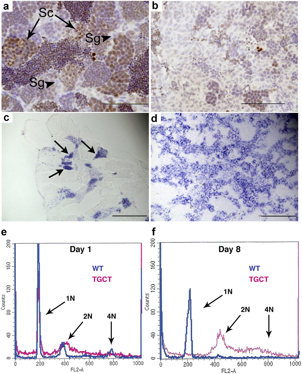
The tgct mutants exhibit impaired differentiation and reduced meiosis in vitro and in vivo. In wild-type testis (A), phosphohistone H2Ax marks clusters of primary spermatocytes undergoing meiosis (arrows). Spermatogonia (arrowheads) are phosphohistone H2AX-negative. The tgct mutant testis (B) exhibits severely reduced clusters of meiotic spermatocytes. The tumor cells are phosphohistone H2AX-negative. (C and D) In situ hybridization for the germ-cell–specific gene ziwi. In wild-type testis (C), ziwi expression is strong in clusters of spermatogonia (arrows) and rapidly declines as cells enter meiosis. The tgct testis tumors (D) show uniformly high expression of ziwi. (E) FACS analysis for DNA content of cultured wild-type and tgct testis on day 1. (F) FACS analysis on day 8 after culturing. Wild-type spermatogonia have completed meiosis to become haploid spermatocytes and spermatozoa (1N), but tgct tumor cells remain diploid (2N) and do not undergo meiosis. [Scale bars: 100 μm (A and B); 200 μm (C and D).]