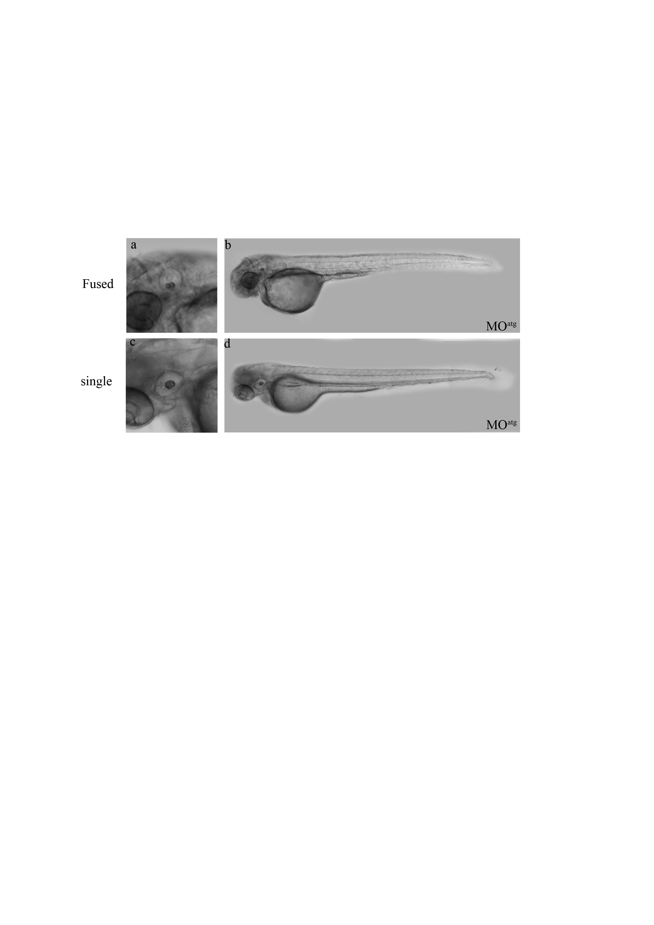
Image


                        Figure Caption


Fig. S3
The morphology of β-tectorin morphants. The ATG MO injected zebrafish embryos with fused (a, b), and single otoliths (c, d) appeared to be normal without obvious defects. All photographs were taken at 72 hpf.
                
                    
                        Acknowledgments


This image is the copyrighted work of the attributed author or publisher, and
ZFIN has permission only to display this image to its users.
Additional permissions should be obtained from the applicable author or publisher of the image.
      Full text @ PLoS One


        