
|
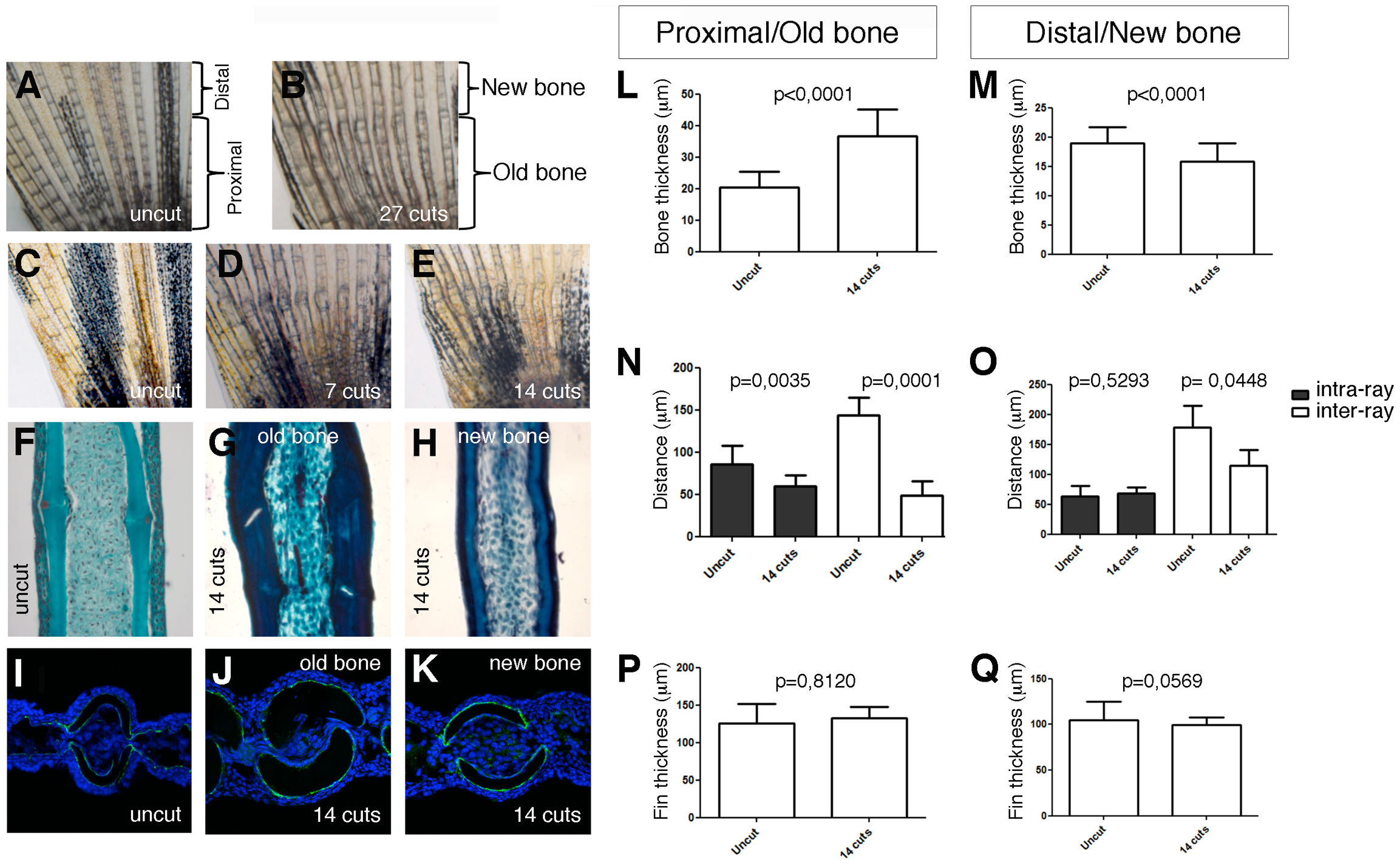
Fig. 4
Consecutive repeated amputations affect the structure of non-regenerate bone.
Picture of the dorsal lobe of an uncut caudal fin (A) and its age-matched sibling after 27 cuts (B). Picture of the dorsal lobe of an uncut caudal fin (C) and a caudal fin after 7 (D) and 14 cuts (E). Masson′s trichrome staining of longitudinal sections of an uncut bony ray (F) and of an old (G) and regenerated (H) regions of a bony ray after 14 cuts. Confocal images of transverse sections of a Zns5 immunostained proximal region of an uncut caudal fin (I) and of the old (J) and new (K) tissue of a caudal fin after 14 cuts. Quantification of the bone thickness, inter- and intra-ray tissue and fin thickness in the old (L, N, P) and new (M, O, Q).