
|
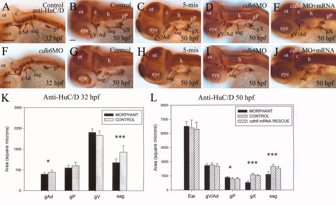
Fig. 3
Development of the cranial and lateral line ganglia requires Cdh6 function. A–J: Anti-HuC/D immunoperoxidase staining of embryos, showing lateral views (anterior to the left and dorsal up) of the head region. K,L: Histograms representing the area/size (square microns; n = 13 for all measured ganglia) of anti-HuC/D labeled cranial and lateral line ganglia, comparing control embryos (gray bars), cdh6 morphants (dark bars) and cdh6 mRNA rescued embryos (bars with diagonal lines). One asterisk indicates significant difference (P = 0.0013), whereas three asterisks indicate highly significant difference (P < 0.001). gX, vagal ganglion; ot, optic tectum. Other abbreviations are the same as in Figure 1. Scale bars = 100 µm in A,F, 50 μm in B–E,G–L.