
|
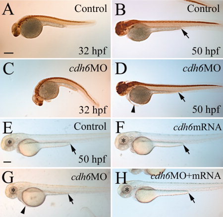
Fig. 2
Overall cdh6 loss-of-function phenotype. Lateral views (anterior to the left and dorsal up) of whole-mount embryos processed for anti-HuC/D immunoperoxidase staining. A–D: The morphants (C,D, injected with cdh6 MO2 [morpholino oligonucleotide 2] showing moderate phenotype) were similar in body and yolk size as uninjected control embryos (A,B), but had smaller head and eyes, curved body at younger (e.g., 32 hours postfertilization) stages, shortened yolk extension and edema in the thorax, as shown in our previous publication (Liu et al., 2008a). E–H: Live embryos raised in PTU (1-phenyl-2-thiourea, 0.003%) -treated fish water. The eyes are indicated by asterisks, edema in the thorax is indicated by an arrowhead, and the end of the yolk extension is indicated by an arrow. Scale bar = 250 μm for all panels.