
|
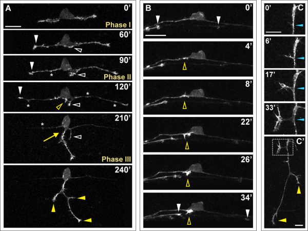
Fig. 4 F-actin distribution in developing RB neurons. F-actin labeled in individual RB neurons by transient mosaic expression of mCh-UtrCH in wild-type embryos. Dorsal-lateral views, anterior is left. Images are confocal projections. (A) Time-lapse showing F-actin distribution during RB neuron development. F-actin is concentrated in the growth cones of central axons (white arrowheads) and peripheral axon branches (yellow arrowheads) as they extend. Accumulation of F-actin occurs at the basal edge of the cell body and becomes concentrated at the distal tip of a neurite that initiates posterior to the cell body and later retracts (white open arrowheads). A second neurite initiates directly off the cell body (yellow open arrowhead) and becomes established as the peripheral axon (yellow arrow). F-actin signal is distributed along the central axon shafts during phase I, but is diminished in the axon shaft during phases II and III. Transient F-actin patches occur along the central axon shafts during phases II and III (some indicated with asterisks) and decrease in frequency over time. See Additional file 3 for movie. (B) Time-lapse showing F-actin accumulation during peripheral axon initiation (open yellow arrowheads). White arrowheads indicate central axon growth cones of neighboring labeled RB neurons extending through the field of view. (C) Time-lapse showing F-actin dynamics during interstitial branch formation in a peripheral axon. F-actin accumulates in a filopodium (0′, 6′) then concentrates in the growth cone of the extending branch (17′, 33′). Blue arrowheads indicate branch point. (C′) Low magnification view of branched peripheral axon at the end of the time-lapse. Box indicates region shown in (C). Yellow arrowheads indicate peripheral growth cones. Time is displayed in minutes. Scale bars in (A,B) = 25 μm, in (C) = 10 μm.