
|
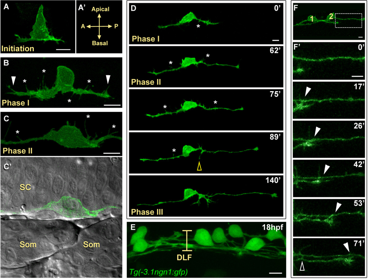
Fig. 1 Central axon development in RB neurons. All images are dorsal-lateral views with anterior to the left and are confocal projections unless noted. Individual RB neurons labeled by transient mosaic expression of TagRFP-CAAX (pseudocolored green) (A-D) or GFP-CAAX (F) in wild-type embryos. (A) Central axon initiation. Central axons form at the basal surface of the cell body at the anterior (A) and posterior (P) sides of the cell. (A′) Indicates orientation. (B) RB neuron in phase I of central axon outgrowth. RB has thick central axons with many filopodial protrusions (asterisks) along the axon shafts and growth cones (arrowheads). (C,C′) RB neuron in phase II of central axon outgrowth. (C) The central axon shafts display transient filopodial protrusions (asterisks). Growth cones have extended out of the field of view. (C′) Single XY plane (green and DIC channel overlay) of RB in (C). The central axons grow along the edge of the spinal cord (SC) neuroepithelium. Som, somites. (D) Selected images from an approximately 3.5-hour time-lapse of RB central axon outgrowth, phases I to III. The peripheral axon initiates during phase II (yellow open arrowhead). Asterisks indicate transient filopodial protrusions during phases I and II. See Additional file 1 for a movie. (E) Image showing all RB neurons labeled in a transgenic Tg(-3.1ngn1:gfp) embryo. At 18 hpf, the dorsal longitudinal fascicle (DLF) is loosely bundled (bracket). (F,F′) Central axon fasciculation. (F) Two labeled RB neurons (1 and 2) have begun to extend central axons. Box indicates area shown in (F′). (F′) Time-lapse of RB central axon fasciculation starting at 17 hpf. RB1 growth cone contacts RB2 axon as it extends (arrowheads) and RB1 axon makes transient lateral filopodial contacts (open arrowhead). Time is displayed in minutes. Scale bars = 10 μm.