
Image
|
Figure Caption
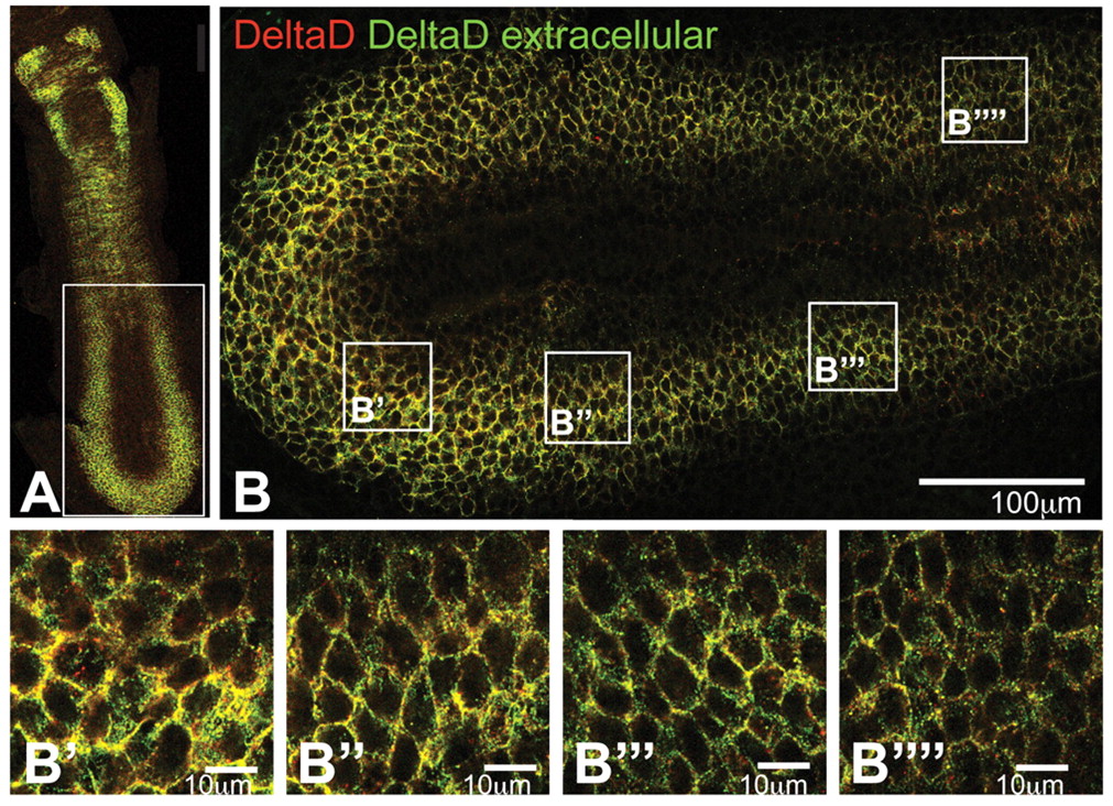
Fig. 8 The subcellular localisation of DeltaD in the PSM of a mib mutant zebrafish embryo. (A-B′′′′) Optical sections of a flat-mount immunostained with zdd2 before (green) and after (red) permeabilisation, as in Fig. 6, to distinguish between intracellular and extracellular DeltaD. The caudorostral gradients in the quantity of DeltaD per cell and in its degree of internalisation, as evident in wild type (see Fig. 6), are lost in the mib mutant, producing a uniform distribution of DeltaD that is at the cell surface throughout the PSM (see B-B′′′′).
Acknowledgments
This image is the copyrighted work of the attributed author or publisher, and
ZFIN has permission only to display this image to its users.
Additional permissions should be obtained from the applicable author or publisher of the image.
Full text @ Development