
|
Fig. 4
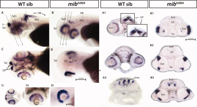
bhlhb5 mRNA expression. A–D: Whole-mounts are shown in lateral (A,B) and dorsal views (C,D). Ci,Di: Smaller panels show a higher magnification of the eye, in lateral views. Note the mibhi904 mutant eye where the retinal layers fail to differentiate. A1–A3: Whole-mount in situ hybridization (WISH) transversal cryostat sections of a stage 72 hours postfertilization (hpf) wild-type (WT). A4–A5: WISH coronal cryostat sections of a stage 72 hpf WT. B1–B3: WISH transversal cryostat sections of a stage 72 hpf mibhi904 mutant. Abbreviations as above: CCe, cerebellum; INL, inner nuclear layer; GCL, ganglion cell layer; ONL, outer nuclear Layer; OP, olfactory pits. Scale bar = 0.2 mm in A–D, 0.1 mm in A1–A5,B1–B3, 0.05 mm in Ci,Di.Figure 5. The hoxa5a and hoxb5b mRNA expression. A,C,E,G: In the upper panels, whole-mounts are shown in lateral views. B,D,F,H: Lower panels are higher magnification and same orientation of area framed in A, C, E, G. Abbreviations as above: Hr, hindbrain rhombomeres; PA, pharyngeal arch. Scale bars = 0.2 mm in A,C,E,G, 0.1 mm in B,D,F,H.