
|
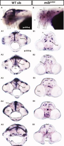
Fig. 2
her4.2 mRNA expression in wild-type (WT) sibling (left panels) and mibhi904 mutant zebrafish (right panels) by in situ hybridization. A,B: Whole-mounts are shown in lateral views. A1–A5: WISH transversal cryostat sections of a stage 72 hr postfertilization (hpf) WT sibling at levels illustrated by the dashed lines in A. B1–B5: WISH transversal cryostat sections of a stage 72 hpf mibhi904 mutant at levels illustrated by the dashed lines in B. Note that dashed lines were indicatively drawn here (and in all subsequent figures) to reflect the approximate cutting angle and location. ac, anterior commissure; DC, diencephalon; DT, dorsal thalamus; EmT, eminentia thalami; H, hypothalamus; HB, hindbrain; Hc, caudal hypothalamus; Hi, intermediate hypothalamus; lfb, lateral forebrain bundle; MO, medulla oblongata; P, pallium; Po, preoptic region; Poc, postoptic commissure; Pr, pretectum; PTd, dorsal part of posterior tuberculum; PTv, ventral part of posterior tuberculum; S, subpallium; T, midbrain tegmentum; Tel, telencephalon; TeO, tectum opticum; VT, ventral thalamus. Scale bar = 0.2 mm in A,B, 0.1 mm in A1–A5,B1–B5.