
|
Fig. S5
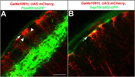
The Position of Radial Glial Endfeet Relative to RGC Axons and Slit2-GFP, Related to Figure 7 Optical sections of a 6 dpf tectum.(A) Radial glia, labeled with Gal4s1061t crossed to UAS:mmCherry (red), and RGC axons, labeled with Pou4f3:mGFP (green). Endfeet (arrow) are in close proximity and slightly superficial to the thin layer of RGC axons in the SO (weak green signal, indicated by arrowheads). The strong green signal in the deeper neuropil corresponds to the two SFGS sublayers, SFGSD and SFGSF, where most Pou4f3:GFP axons are projecting.(B) Radial glia, labeled with Gal4s1061t crossed to UAS:mmCherry (red) and heatshock-induced Slit2-GFP (green). The Slit2-GFP signal coincides with, and appears to be enriched around, the glial endfeet (arrow).
Reprinted from Cell, 146(1), Xiao, T., Staub, W., Robles, E., Gosse, N.J., Cole, G.J., and Baier, H., Assembly of Lamina-Specific Neuronal Connections by Slit Bound to Type IV Collagen, 164-176, Copyright (2011) with permission from Elsevier. Full text @ Cell