
|
Fig. 6
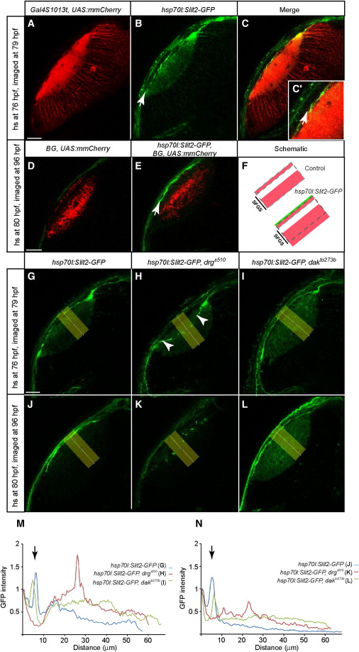
Slit Localization in the Optic Tectum Requires Col4a5 but Not HSPGs(A–C) Optical section of a 79 hpf hsp70l:Slit2-GFP, Gal4s1013t, and UAS:mmCherry tectum, heat shocked at 76 hpf. Many tectal neurons are labeled with mmCherry (red, A). Slit2-GFP (green, B) is enriched at the surface of the tectal neuropil (arrow). (C) is a merged image.(D–F) Optical sections of 96 hpf hsp70l:Slit2-GFP, Pou4f3:Gal4 (Brn3c:Gal4, BG), and UAS:mmCherry tecta, heat shocked at 80 hpf. RGC axons (red) are shown in non-heat-shocked (D) and heat-shocked fish (E). Arrow points at the accumulation of Slit2-GFP superficial to SO. (F) is a schematic summary.(G–L) Optical sections of hsp70l:Slit2-GFP tecta in the WT (G and J), drgs510 (H and K), and dak t0273b (I and L). Larvae were either heat shocked at 76 hpf and imaged at 79 hpf (G–I) or heat shocked at 80 hpf and imaged at 96 hpf (J–L).(M and N) Densitometric plots of GFP intensity in the yellow rectangle areas in (G)–(L). (M) shows distribution 3 hr after induction. N shows distribution 16 hr after induction. Intensity is normalized to skin autofluorescence level (first peak at 0 μm). In the WT, Slit2-GFP accumulates beneath the skin (second peak at ~8 µm from the surface, indicated by arrows) and is cleared from the neuropil. In drgs510, Slit2-GFP accumulates in the neuropil, away from the surface. In dak t0273b, Slit2-GFP is surface-localized, but remains also in the neuropil.Scale bars represent 20 μm. See also Figure S4.
Reprinted from Cell, 146(1), Xiao, T., Staub, W., Robles, E., Gosse, N.J., Cole, G.J., and Baier, H., Assembly of Lamina-Specific Neuronal Connections by Slit Bound to Type IV Collagen, 164-176, Copyright (2011) with permission from Elsevier. Full text @ Cell