
|
Fig. S4
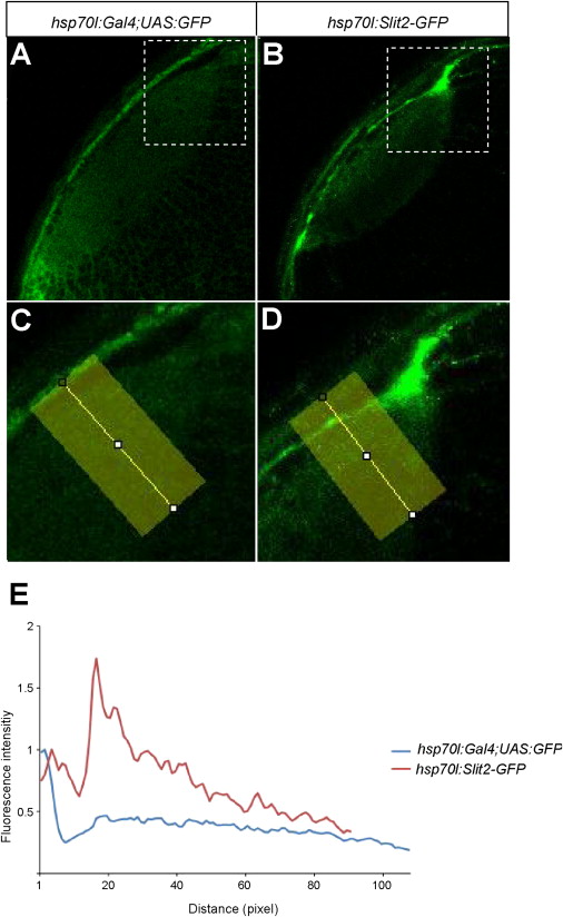
Heat Shock-Induced GFP Expression Is Spatially Uniform, Related to Figure 6(A–D) Optical sections of the tectum in 3 dpf embryos. Genotypes as indicated in the panels. C and D shows higher magnifications of the boxed regions in A and B, respectively.(E) Densitometric plots of GFP intensity from the areas indicated by yellow rectangles. Intensity is normalized to skin autofluorescence level (first peak at about 5 pixels from surface). While GFP is evenly distributed in the neuropil after induction, Slit2-GFP accumulates beneath the skin (second peak at 18 pixels of the red trace). Ten pixels correspond to ca. 5 μm.
Reprinted from Cell, 146(1), Xiao, T., Staub, W., Robles, E., Gosse, N.J., Cole, G.J., and Baier, H., Assembly of Lamina-Specific Neuronal Connections by Slit Bound to Type IV Collagen, 164-176, Copyright (2011) with permission from Elsevier. Full text @ Cell