
|
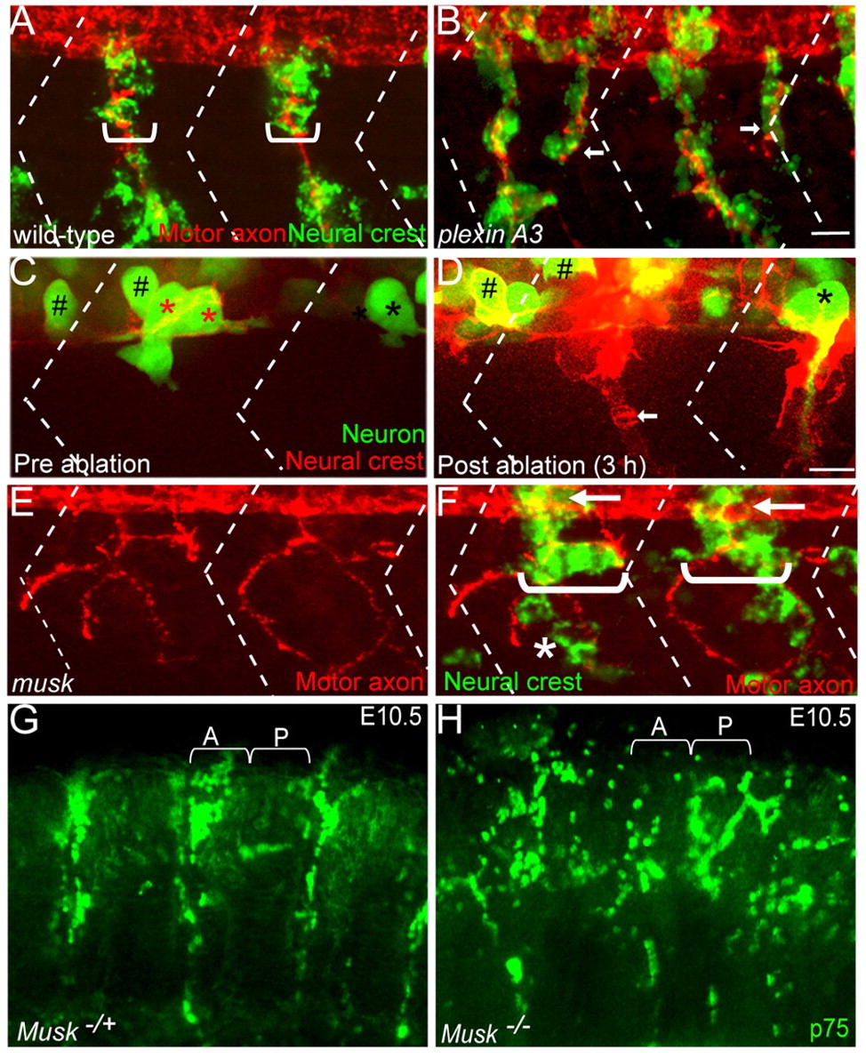
Fig. 2
The role of motor axons and MuSK signaling during segmental neural crest cell migration. (A,B) Lateral views of 28 hpf wild-type (A) and plexin A3 mutant (B) zebrafish embryos with neural crest cells in green (crestin) and motor axons in red (znp-1/SV2). Brackets indicate width of neural crest cell stream. (C,D) Lateral views of a double Tg(mnx1:GFP)ml2; (sox10:mRFP)vu234 embryo expressing GFP (green) in motor neurons (asterisks) and in interneurons (hash marks), and mRFP (red) in neural crest cells before laser ablation (C) and three hours following motor neuron ablation (D; arrow points to ventral migration of neural crest cells). Red asterisks in C indicate ablated motor neurons. (E,F) Lateral views of a 28 hpf musk mutant embryo stained to reveal neural crest cells (crestin, green) and motor axons (znp1+SV2, red). Arrows point to migrating neural crest cells before they join the path shared with motor axons. In the musk mutant embryo, neural crest cells and motor axons deviate from their central path and migrate over a wider region of the somite (compare brackets in F with A; asterisk marks neural crest cells taking a path independent of motor axons). Dashed lines in A-F indicate approximate positions of somite boundaries. Scale bars: 10 μm. (G,H) Lateral views of whole-mount E10.5 mouse embryos stained with p75 antibody labeling neural crest cells. Neural crest cells migrate through the anterior sclerotome in the wild-type sibling embryo (A), but migrate non-segmentally in the Musk knockout embryo. A, anterior somite; P, posterior somites.