
|
Fig. 9
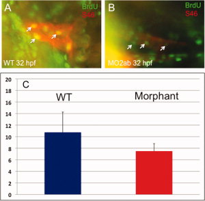
Cardiomyocyte proliferation is not decreased in the atrium of tbx2ab morphants. Embryos were pulse-labeled with bromodeoxyuridine (BrdU), and after being fixed, were co-stained to detect BrdU+ cells (green) and S46+ atrial cardiomyocytes (red). Hearts were imaged and the yellow (double positive) cells counted (examples indicated by the small arrows). A,B: The top panels show representative wild-type (A) and morphant (B) embryos at <32 hpf. C: The lower panel shows quantification of the average BrdU+ cardiomyocytes, in each case from several randomly chosen embryos, n = 4. According to Student′s t-test, P = 0.13. The result is consistent with the fact that the atrium is not altered in cell numbers.