
|
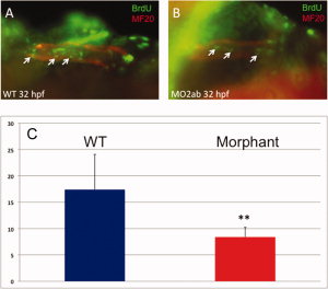
Fig. 8
Cardiomyocyte proliferation is decreased in the tbx2ab morphants. Embryos were pulse-labeled with bromodeoxyuridine (BrdU), and after being fixed, were co-stained to detect BrdU+ cells (green) and MF20+ cardiomyocytes (red). Hearts were imaged, and the yellow (double positive) cells counted (examples indicated by the small arrows). A,B: The top panels show representative wild-type (A) and morphant (B) embryos at <32 hpf. C: The lower panel shows quantification of the average BrdU+ cardiomyocytes, in each case from several randomly chosen embryos, n = 5. The ** indicates statistical significance compared with wild-type, according to Student′s t-test, P < 0.02. This experiment was repeated several times and reproducibly showed significantly decreased relative levels of BrdU+ cardiomyocytes in morphants, although the actual number of double-labeled cells varies depending on the efficiency of BrdU labeling.