
|
Fig. 2
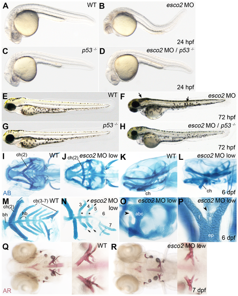
Craniofacial development is abnormal in esco2 morphants.
A–D, At 24 hpf, esco2 morphants (B) had a smaller head, smaller eyes and noticeable cell death in the head region compared to wild type (A), and could be partially rescued by depletion of p53 (p53 -/-, C, D). E–H, At 72 hpf esco2 morphants lacked a jaw and showed an abnormal pigmentation pattern (arrows) (F compared with E, wild type). The phenotype was enhanced by depletion of p53 (G, H). A–H, lateral views of live larvae, anterior to the left. I–P, Craniofacial cartilage was stained with Alcian Blue (AB) at 6 dpf. A low dose of esco2 MO was injected to allow for cartilage development (esco2 MO low). Numbers of the pharyngeal arch elements are indicated (2–7). I, J, Ventral views. K, L, lateral views. Pharyngeal arch elements were malformed or missing in esco2 morphants (J, L) compared to wild type (I, K). M, N, closer views of dissected pharyngeal arch elements in wild type (M) and esco2 morphant (N). The arrows point to the kinks in arches 3 and 4. The asterisks mark elongated hypobranchials (hb) in an esco2 morphant. O, closer view of anterior basicranial commissure (abc) in an esco2 morphant, where a gap in the structure was observed (arrowhead). P, closer view of the neurocranium with additional cell structures (arrowhead) at the base of the trabeculae (t) in an esco2 morphant. Q, R, Alizarin Red (AR) staining of bone matrix at 7 dpf (ventral views) revealed that ossification is normal in an esco2 morphant (R, compare to wild type in Q). The close-ups show branchial arch 7. ch, ceratohyal; bh, basihyal; hb, hypobranchial; cb, ceratobranchial; abc, anterior basicranial commissure; t, trabeculae; ep, ethmoid plate.