
Image
|
Figure Caption
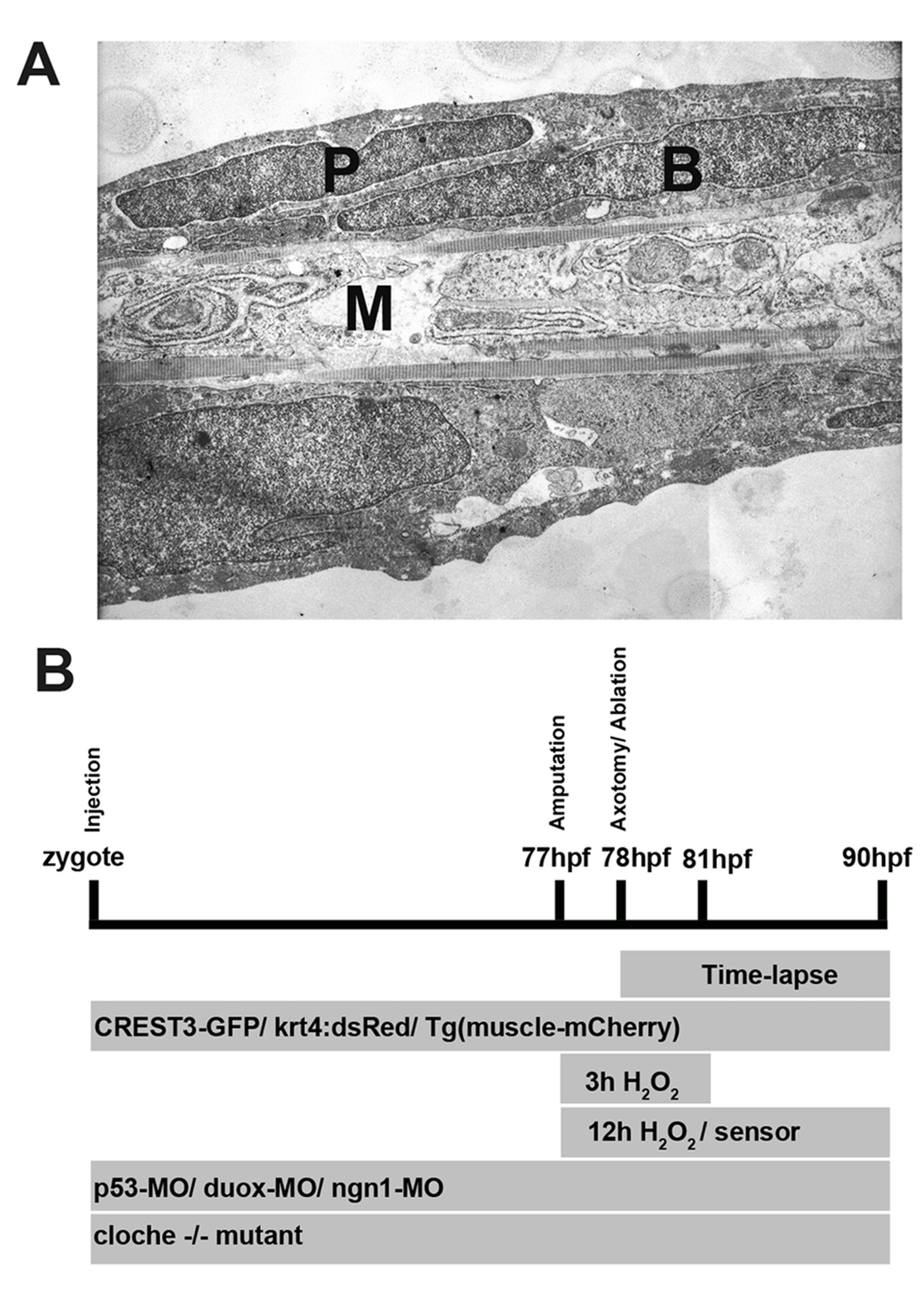
Fig. S1
Ultrastructure of a larval fin and experimental design. (A) Transmission electron micrograph of a sagittal section through the caudal fin at 48 hpf. The skin consists of two cell layers, the outer periderm (P) and inner epidermal basal cells (B), which are separated by a basement membrane from medially located muscle (M). Magnification is 4,800×. (B) Timeline of experimental procedures. hpf, hours post fertilization.
Acknowledgments
This image is the copyrighted work of the attributed author or publisher, and
ZFIN has permission only to display this image to its users.
Additional permissions should be obtained from the applicable author or publisher of the image.
Full text @ PLoS Biol.